
Page 278 - The Veterinary Laboratory and Field Manual 3rd Edition
P. 278
Microbiology 247
Initial growth of cells is done in various types samples using filtration to remove bacteria and
of cell culture flasks, disposable glass flasks and other microorganisms). After a short period of
polyethylene flasks of various sizes are commonly incubation (≈30–60 min) to allow attachment
used. The main applications of cell culture are of the virus to the cells, the fluid is poured off
to grow viruses to high titres, to identify viruses and replaced. Cell culture media usually con-
based on the cytopathic effects induced in cell tains a red dye that changes colour (due to the
culture, and for the quantification of replicating change of pH) when the media needs replacing.
viruses in samples using plaque assays (Figure Following a period of incubation (≈2–5 days)
4.23). However, some viruses are more difficult with the virus, necrotic cells may be observed
to culture than others. The correct cell line and due to virus growth within the cells (Figure
the ideal environmental conditions required can 4.24). The extent of necrosis (cell death dem-
only be selected when the sample submitter has onstrated by shrunken nuclei and withering cell
provided a clear list of diagnostic differentials in margins or plaque formation) can be noted and
advance. For these procedures, establishment of recorded to determine the relative number of
monolayers of cells in six-, twelve- and twenty- viral particles present in the virus inoculum. In
four-well plates is generally required. general, it is assumed that each area of necrosis
Once a good monolayer of cells has been or plaque formation results from infection by
produced, the media used to grow the cells can one viral particle.
be replaced with nutrient media containing the
virus of interest (this can be extracted from
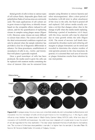
Figure 4.23 In plaque assay the virus inoculum is ten-fold serially diluted in phosphate buffered saline and
inoculated into the monolayer of cells which are permissive to the inoculating virus. In this figure, avian
influenza virus titration has been done in Madin-Darby Canine Kidney (MDCK) cells. After two days of
inoculation of the MDCK cell monolayer, the cells have been stained with crystal violet to see the extent
of cell damage due to the virus replication. The clear areas represent the loss of cells due to viral replica-
tion. The number of infectious virus particles in sample 1 is higher than the sample 2. See also Plate 15.
Source: M. Sarjoon Abdul-Cader, University of Calgary, Canada.
Vet Lab.indb 247 26/03/2019 10:25