
Page 275 - The Veterinary Laboratory and Field Manual 3rd Edition
P. 275
244 Susan C. Cork and Roy Halliwell
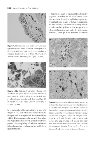
Techniques such as immunohistochemistry
(Figure 4.19a and b) and the use of special stains
have also been devised to highlight the presence
of viral antigens in cell or tissue preparations.
As with bacteria, differential staining makes
it easier to distinguish the viral antigens from
other small particles seen under the microscope.
However, although it is possible to resolve
(a)
Figure 4.18a Canine airway epithelium with intra-
cytoplasmic inclusions of canine distemper virus
(the arrow indicates eosinophilic intracytoplasmic
inclusion bodies). See also Plate 11. Photo: Dr
Jennifer Davies, University of Calgary, Canada.
(b)
Figure 4.18b Trachea of a chicken infected with
infectious laryngotracheitis virus with multinucle-
ated syncytial cell formation (the arrow indicates
a multinucleated syncytial cell). See also Plate 12.
Photo: Dr M. Faizal Abdul-Careem, University of Figure 4.19 Immunohistochemistry staining can be
Calgary, Canada. performed in frozen sections or formalized sections
to visualize viral antigens. (a) A frozen section of
be evidence of viral inclusion bodies in host cells Bursa of Fabricius of a chicken infected with Marek’s
(Figure 4.18a) and other virus induced cellular disease virus (the arrow indicates the brown colour
changes such as syncytial cell formation (Figure stained viral antigens). Photo: Dr Shayan Sharif,
4.18b). The appearance of these will depend on University of Guelph, Canada. (b) A formalized brain
the stage of infection as well as the type of virus section of a dog infected with canine distemper
present. The morphology of inclusion bodies and virus (the arrow indicates the brown colour stained
other changes can be characteristic and help to viral antigens). See also Plate 13a & b. Photo: Dr
identify the virus. Cameron Knight, University of Calgary, Canada.
Vet Lab.indb 244 26/03/2019 10:25