
Page 276 - The Veterinary Laboratory and Field Manual 3rd Edition
P. 276
Microbiology 245
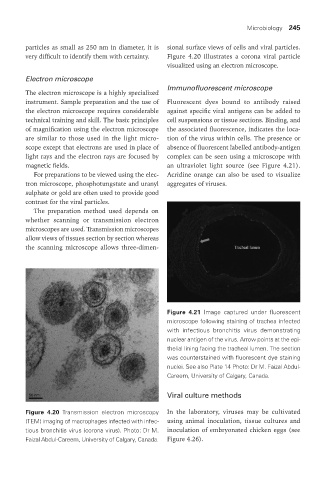
particles as small as 250 nm in diameter, it is sional surface views of cells and viral particles.
very difficult to identify them with certainty. Figure 4.20 illustrates a corona viral particle
visualized using an electron microscope.
Electron microscope
Immunofluorescent microscope
The electron microscope is a highly specialized
instrument. Sample preparation and the use of Fluorescent dyes bound to antibody raised
the electron microscope requires considerable against specific viral antigens can be added to
technical training and skill. The basic principles cell suspensions or tissue sections. Binding, and
of magnification using the electron microscope the associated fluorescence, indicates the loca-
are similar to those used in the light micro- tion of the virus within cells. The presence or
scope except that electrons are used in place of absence of fluorescent labelled antibody-antigen
light rays and the electron rays are focused by complex can be seen using a microscope with
magnetic fields. an ultraviolet light source (see Figure 4.21).
For preparations to be viewed using the elec- Acridine orange can also be used to visualize
tron microscope, phosphotungstate and uranyl aggregates of viruses.
sulphate or gold are often used to provide good
contrast for the viral particles.
The preparation method used depends on
whether scanning or transmission electron
microscopes are used. Transmission microscopes
allow views of tissues section by section whereas
the scanning microscope allows three-dimen-
Figure 4.21 Image captured under fluorescent
microscope following staining of trachea infected
with infectious bronchitis virus demonstrating
nuclear antigen of the virus. Arrow points at the epi-
thelial lining facing the tracheal lumen. The section
was counterstained with fluorescent dye staining
nuclei. See also Plate 14 Photo: Dr M. Faizal Abdul-
Careem, University of Calgary, Canada.
viral culture methods
Figure 4.20 Transmission electron microscopy In the laboratory, viruses may be cultivated
(TEM) imaging of macrophages infected with infec- using animal inoculation, tissue cultures and
tious bronchitis virus (corona virus). Photo: Dr M. inoculation of embryonated chicken eggs (see
Faizal Abdul-Careem, University of Calgary, Canada. Figure 4.26).
Vet Lab.indb 245 26/03/2019 10:25