
Page 423 - The Veterinary Laboratory and Field Manual 3rd Edition
P. 423
A B
C D
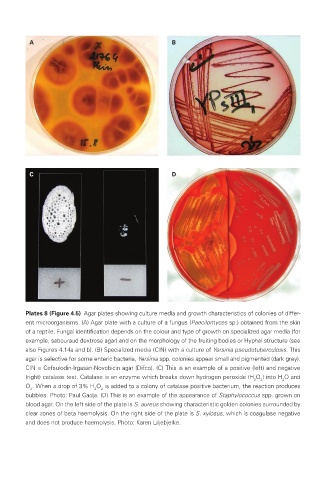
Plates 8 (Figure 4.5) Agar plates showing culture media and growth characteristics of colonies of differ-
ent microorganisms. (A) Agar plate with a culture of a fungus (Paecilomyces sp.) obtained from the skin
of a reptile. Fungal identification depends on the colour and type of growth on specialized agar media (for
example, sabouraud dextrose agar) and on the morphology of the fruiting bodies or Hyphal structure (see
also Figures 4.14a and b). (B) Specialized media (CIN) with a culture of Yersinia pseudotuberculosis. This
agar is selective for some enteric bacteria, Yersinia spp. colonies appear small and pigmented (dark grey).
CIN = Cefsulodin-Irgasan-Novobicin agar (Difco). (C) This is an example of a positive (left) and negative
(right) catalase test. Catalase is an enzyme which breaks down hydrogen peroxide (H O ) into H O and
2 2 2
O . When a drop of 3% H O is added to a colony of catalase positive bacterium, the reaction produces
2 2 2
bubbles. Photo: Paul Gadja. (D) This is an example of the appearance of Staphylococcus spp. grown on
blood agar. On the left side of the plate is S. aureus showing characteristic golden colonies surrounded by
clear zones of beta haemolysis. On the right side of the plate is S. xylosus, which is coagulase negative
and does not produce haemolysis. Photo: Karen Liljebjelke.
Veterinary-plates.indd 6 26/03/2019 10:14