
Page 427 - The Veterinary Laboratory and Field Manual 3rd Edition
P. 427
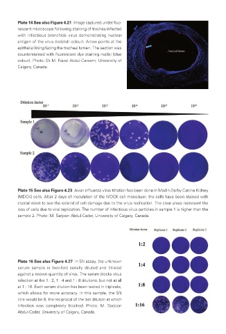
Plate 14 See also Figure 4.21  Image captured under fluo-
            rescent microscope following staining of trachea infected
            with infectious bronchitis virus demonstrating nuclear
            antigen of the virus (reddish colour). Arrow points at the
            epithelial lining facing the tracheal lumen. The section was
            counterstained with fluorescent dye staining nuclei (blue
            colour). Photo: Dr M. Faizal Abdul-Careem, University of
            Calgary, Canada.


            Plate 15 See also Figure 4.23  Avian influenza virus titration has been done in Madin-Darby Canine Kidney
            (MDCK) cells. After 2 days of inoculation of the MDCK cell monolayer, the cells have been stained with
            crystal violet to see the extend of cell damage due to the virus replication. The clear areas represent the
            loss of cells due to viral replication. The number of infectious virus particles in sample 1 is higher than the
            sample 2. Photo: M. Sarjoon Abdul-Cader, University of Calgary, Canada.


            Plate 16 See also Figure 4.27  In SN assay, the unknown
            serum sample is two-fold serially diluted and titrated
            against a known quantity of virus. The serum blocks virus
            infection at the 1 : 2, 1 : 4 and 1 : 8 dilutions, but not at all
            at 1 : 16. Each serum dilution has been tested in triplicate,
            which allows for more accuracy. In this sample, the SN
            titre would be 8, the reciprocal of the last dilution at which
            infection was completely blocked. Photo: M. Sarjoon
            Abdul-Cader, University of Calgary, Canada.


       Veterinary-plates.indd   10                                                         26/03/2019   10:14