
Page 432 - The Veterinary Laboratory and Field Manual 3rd Edition
P. 432
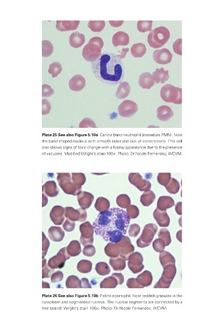
Plate 25 See also Figure 5.10a  Canine band neutrophil (immature PMN). Note
                          the band shaped nucleus with smooth sides and lack of constrictions. This cell
                          also shows signs of toxic change with a foamy appearance due to the presence
                          of vacuoles. Modified Wright’s stain 100×. Photo: Dr Nicole Fernandez, WCVM.


                          Plate 26 See also Figure 5.10b  Feline eosinophil. Note reddish granules in the
                          cytoplasm and segmented nucleus. The nuclear segments are connected by a
                          fine strand. Wright’s stain 100×. Photo: Dr Nicole Fernandez, WCVM.


       Veterinary-plates.indd   15                                                         26/03/2019   10:14