
Page 435 - The Veterinary Laboratory and Field Manual 3rd Edition
P. 435
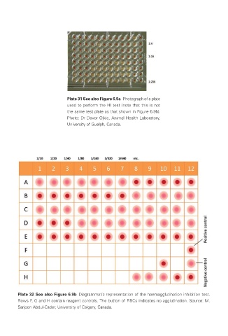
Plate 31 See also Figure 6.9a  Photograph of a plate
                                used to perform the HI test (note that this is not
                                the same test plate as that shown in Figure 6.9b).
                                Photo: Dr Davor Ojkic, Animal Health Laboratory,
                                University of Guelph, Canada.


            Plate 32 See also Figure 6.9b  Diagrammatic representation of the haemagglutination inhibition test.
            Rows F, G and H contain reagent controls. The button of RBCs indicates no agglutination. Source: M.
            Sarjoon Abdul-Cader, University of Calgary, Canada.


       Veterinary-plates.indd   18                                                         26/03/2019   10:14