
Page 426 - The Veterinary Laboratory and Field Manual 3rd Edition
P. 426
A
B
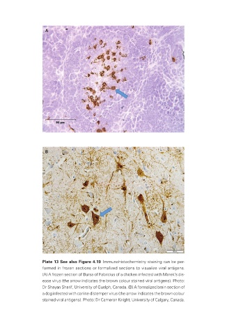
Plate 13 See also Figure 4.19 Immunohistochemistry staining can be per-
formed in frozen sections or formalized sections to visualize viral antigens.
(A) A frozen section of Bursa of Fabricius of a chicken infected with Marek’s dis-
ease virus (the arrow indicates the brown colour stained viral antigens). Photo:
Dr Shayan Sharif, University of Guelph, Canada. (B) A formalized brain section of
a dog infected with canine distemper virus (the arrow indicates the brown colour
stained viral antigens). Photo: Dr Cameron Knight, University of Calgary, Canada.
Veterinary-plates.indd 9 26/03/2019 10:14