
Page 448 - The Veterinary Laboratory and Field Manual 3rd Edition
P. 448
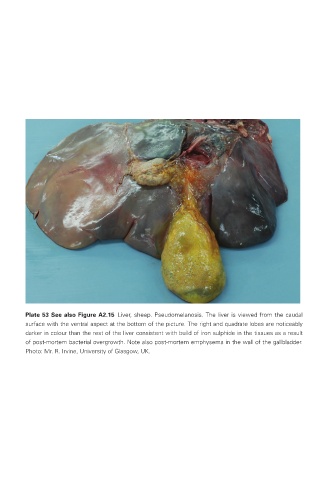
Plate 53 See also Figure A2.15  Liver, sheep. Pseudomelanosis. The liver is viewed from the caudal
                surface with the ventral aspect at the bottom of the picture. The right and quadrate lobes are noticeably
                darker in colour than the rest of the liver consistent with build of iron sulphide in the tissues as a result
                of post-mortem bacterial overgrowth. Note also post-mortem emphysema in the wall of the gallbladder.
                Photo: Mr. R. Irvine, University of Glasgow, UK.


       Veterinary-plates.indd   31                                                         26/03/2019   10:14