
Page 1071 - Adams and Stashak's Lameness in Horses, 7th Edition
P. 1071
Lameness in the Young Horse 1037
trabecular bone, making it incapable of correcting an This is partly why joint luxations appear to be rare in
ALD. The timing of the physeal closure depends on the young horses.
VetBooks.ir open for several years (Table 10.1). However, it is impor
specific bone. Some close early in life and others remain
tant to note that functional (remaining growth potential)
physeal closure occurs well before radiographic closure, CLASSIFICATION AND TREATMENT OF PHYSEAL
INJURIES/FRACTURES
and this difference has important bearing on the timing
of surgery used to correct ALDs. In general, the further The most widely accepted classification of growth
55
distal on the limb the physis is located, the sooner longi plate injuries is based on the Salter‐Harris system, which
tudinal growth will become functionally inactive. For separates the injuries into six specific types (Figure 10.5).
47
instance, the distal metacarpal/metatarsal physes close The system was designed for humans but has been
sooner compared with the distal radial or distal tibial applied to horses to permit equine clinicians to commu
physes (Table 10.1), making correction of ALDs of the nicate effectively when describing such injuries.
fetlock more important early in life than those of the car Physeal fractures are relatively common in foals
pus or tarsus. In addition, any injury to the physis such (accounting for approximately 20% of fractures) and
19
as excessive pressure, direct trauma, traction, circulatory may be considered more serious than diaphyseal frac
loss, or shearing forces can lead to premature cessation tures because of the risk of disturbed limb growth and
of growth or asynchronous growth. 4,62 Comparatively, articular involvement. 16,17,42 Type I and II physeal frac
circumferential growth of the epiphysis continues uni tures are most common in horses. Frequent locations of
formly regardless of anatomical location until the horse physeal fractures include the distal metacarpus/metatar
is fully mature. 30 sus, distal and proximal radius, proximal humerus,
proximal tibia, and distal and proximal femur
External Trauma to the Physis (Figures 10.6 and 10.7).
These fractures tend to heal quickly but usually
When excessive force is applied to a joint and its reduce the growth potential remaining in the physis via
nearby physis, an epiphyseal or physeal injury is likely to premature closure of the physis at time of injury or dur
occur because the cartilaginous growth plate is weaker ing fracture repair. This can lead to premature limb
than the surrounding bone, ligamentous structures, and shortening or ALD. Certain locations (distal metacar
joint capsule. Physeal and epiphyseal injuries are com pus/metatarsus and proximal tibia) have sparse soft tis
mon in foals. 16,17,42 Injuries that would normally produce sue coverage and can lead to open fractures and
a ruptured ligament or joint dislocation in an adult may increased risk of implant‐associated infection with sur
produce traumatic separation of the physis in a young gical repair. Concurrent injuries to the surrounding
animal. For example, trauma to the fetlock region might synovial, ligamentous, and tendinous structures can
cause a fetlock luxation in an adult horse, whereas a foal also influence overall prognosis. As with any fracture,
might sustain a distal metacarpal/tarsal physeal fracture. the greater the degree of instability, displacement, and
55
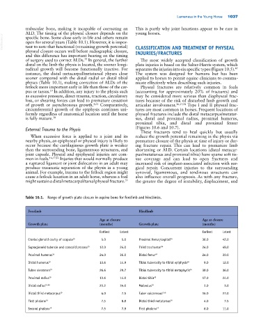
Table 10.1. Range of growth plate closure in equine bone for forelimb and hindlimbs.
Forelimb Hindlimb
Age at closure Age at closure
Growth plate (months) Growth plate (months)
Earliest Latest Earliest Latest
Cranial glenoid cavity of scapula 15 5.0 5.0 Proximal femur/capital 54 36.0 42.0
Supraglenoid tubercle and coracoid process 15 12.0 24.0 Third trochanter 54 24.0 48.0
Proximal humerus 15 24.0 36.0 Distal femur 64 24.0 30.0
Distal humerus 19 13.6 14.9 Tibial tuberosity to tibial epiphysis 64 9.0 12.0
Tuber olecranon 19 26.6 29.7 Tibial tuberosity to tibial metaphysis 64 30.0 36.0
Proximal radius 19 13.6 14.0 Distal tibia 65 17.0 24.0
Distal radius 19,56 23.2 35.0 Malleolus 10 3.0 3.0
Distal third metacarpus 19 4.0 7.5 Tuber calcaneous 2,56 16.0 27.0
First phalanx 19 7.5 8.8 Distal third metatarsus 19 4.0 7.5
Second phalanx 19 7.5 7.9 First phalanx 19 8.0 11.0