
Page 1076 - Adams and Stashak's Lameness in Horses, 7th Edition
P. 1076
1042 Chapter 10
Trauma to the metaphyseal or epiphyseal growth plates
may also contribute to altered growth, subchondral bone
VetBooks.ir physeal vasculature, all of which may predispose to DOD
damage, avulsion of defective bone, and disruption of the
conditions. Weight, limb conformation, and excessive
exercise may be contributing factors. 50, 57 Genetics most
likely plays a role in the occurrence of these diseases, but
its contribution is difficult to determine. OC is most likely
a polygenic trait with a complex method of inheritance.
66
A genetic predisposition has been demonstrated in dogs,
pigs, and horses. Populations of risk alleles from two
49
separate populations of OC(D) affected Standardbreds
suggested that there is a true risk locus as a contributing
36
etiology. However, in most cases, the underlying cause of
the DOD condition is multifactorial, usually obscure, and
often never determined. 4,62 Factors that may have contrib
uted to the disease process are often long gone by the time
a veterinarian is asked to evaluate the horse. In addition,
the timing at which risk factors may exert their effects on
bone growth and development is currently unknown.
Discussions of each specific DOD condition seen in grow
ing horses are presented throughout this chapter.
EPIPHYSITIS/PHYSITIS/PHYSEAL DYSPLASIA
Physitis or epiphysitis is an important generalized
bone disease of young growing horses characterized by
inflammation and enlargement of the physeal region of
certain immature long bones (Figure 10.9). Most cases
4,5
of physitis occur in young, rapidly growing horses such
as weanlings, with a peak incidence between 4 and
61
8 months of age, but even yearlings and 2‐year‐old
horses may also develop the condition. It may affect a
single or multiple growth plates, but is often bilaterally
symmetrical.
Physeal dysplasia may be a more appropriate term
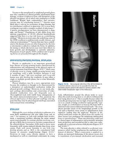
because the condition is thought to be characterized by Figure 10.10. Varus angular deformity of the left front fetlock in
a foal that suffered a fracture on the opposite limb. Note the
a disruption of endochondral ossification within the secondary physitis present with sclerosis (arrows) present in the
physeal growth cartilage. However, in one study, phy distal medial metaphyseal region of the metacarpus.
5
seal cartilage abnormalities and compromise of endo
chondral ossification were not frequently seen in
Thoroughbred foals with visible bony enlargements of foals, inflammation around the physis tends to occur
the distal metacarpus/metatarsus. This questions the when this newly forming bone is not able to withstand the
23
clinical significance of these physeal swellings and sug load being placed on it. This is usually because either the
5
gests that they may be physiological swellings associated normally growing bone has too high of loads being placed
with normal bone remodeling. 23 on it as a result of being overfed for rapid growth, exer
4
cise, weight, or conformation or because the bone itself is
abnormal and cannot withstand normal loads. The meta
5
ETIOLOGY physis provides much of the longitudinal growth in the
foal and is therefore more prone to inflammation from
Although the exact etiology of physitis is unknown, it is repetitive loading than the epiphysis. Regions of dis
5
most likely multifactorial and may differ from case to turbed ossification within the physis from any number of
case. For instance, in foals with multiple limb involve these factors may predispose the underlying subchondral
4,5
ment, a nutritional problem affecting the entire animal bone to microfractures. These microfractures could lead
61
seems most plausible. In contrast, physitis involving a sin to clinical signs of inflammation and potentially stimulate
gle site is likely due to trauma or excessive compression of bone production and remodeling that is often seen radio
the affected physis. Secondary ALDs tend to occur more graphically as sclerosis in horses with physitis (Figures 10.9
4
frequently with trauma‐induced physitis than from other and 10.10). 4,61
causes. Foals with severe lameness in one limb may The term physitis is often referred to as physeal com
develop physitis and an ALD in the contralateral limb pression, which further emphasizes the mechanical com
because of excessive weight‐bearing (Figure 10.10). ponent of physitis. When compression is applied to a
3
55
However, in many cases, physitis appears to have a physis, an increased thickening of the physis occurs due
mechanical as well as a nutritional component. In young to retardation of provisional calcification and increased