
Page 1078 - Adams and Stashak's Lameness in Horses, 7th Edition
P. 1078
VetBooks.ir
A B
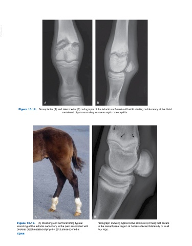
Figure 10.12. Dorsoplantar (A) and lateromedial (B) radiographs of the fetlock in a 2‐week‐old foal illustrating radiolucency at the distal
metatarsal physis secondary to severe septic osteomyelitis.
A B
Figure 10.13. (A) Weanling colt demonstrating typical radiograph showing typical bone sclerosis (arrows) that occurs
knuckling of the fetlocks secondary to the pain associated with in the metaphyseal region of horses affected bilaterally or in all
bilateral distal metatarsal physitis. (B) Lateral‐to‐medial four legs.
1044