
Page 1073 - Adams and Stashak's Lameness in Horses, 7th Edition
P. 1073
Lameness in the Young Horse 1039
VetBooks.ir
A B
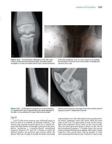
Figure 10.6. (A) Dorsopalmar radiograph of a foal with a type while under anesthesia. There is a type I fracture of the proximal
I fracture of the proximal first phalanx physis. (B) Ventral–dorsal femoral physis. The arrows point out the location of the epiphysis;
radiograph of a foal’s pelvis taken with the foal in dorsal recumbency normal is on the right.
A B
Figure 10.7. (A) Dorsoplantar radiograph of the distal metatarsus fracture. In most situations, these types of fractures should be repaired
of a weanling with a type II physeal fracture. (B) Lateral radiograph of surgically to prevent malalignment of the limb.
the stifle of a long weanling with a type II distal femoral physeal
Type II
and bending forces. The distal third metacarpal/metatar
Type II is the most common type of physeal injury in sal physis commonly incurs this injury when the mare
foals as well as in virtually all domestic animals. The steps on the foal. The periosteum is torn on the convex
55
fracture line extends along the physis for a variable dis side of the angulation but is intact on the concave side.
tance and then breaks out through a portion of the meta Thus, the intact periosteal hinge is always on the side of
physis, producing a triangular‐shaped metaphyseal the metaphyseal fragment. A similar injury also occurs
47
fragment (Figures 10.5 and 10.7). Similar to nearly all in the proximal tibial physis in slightly older foals. Closed
physeal injuries, the germinal cells remain within the reduction with casting alone may be possible in foals
physis. This type of injury is usually the result of shearing with metacarpal/metatarsal type II physeal injuries, but