
Page 1077 - Adams and Stashak's Lameness in Horses, 7th Edition
P. 1077
Lameness in the Young Horse 1043
survival of chondrocytes. 4,55 This is an autocorrection CLINICAL SIGNS
phenomenon in which the animal corrects any minor The clinical signs in young foals are variable but usually
VetBooks.ir are continually under challenge in the growing animal by include a stiff gait with a shortened cranial phase to the
angulation of the limb axis. However, loads on the physis
stride, increased amount of time lying down, and trem
the increase in both body weight and activity level. If the
6
compression is beyond the physiologic limits of the bling when standing, potentially buckling forward at the
5,59,61,63
physis, complete arrest of endochondral ossification may carpus or fetlock (Figure 10.13). In general, physitis
occur. The end result is asynchronous physeal growth occurs more commonly in distal versus proximal physes,
and the development of an ALD together with physitis. as distal growth plates have less soft tissue support and are
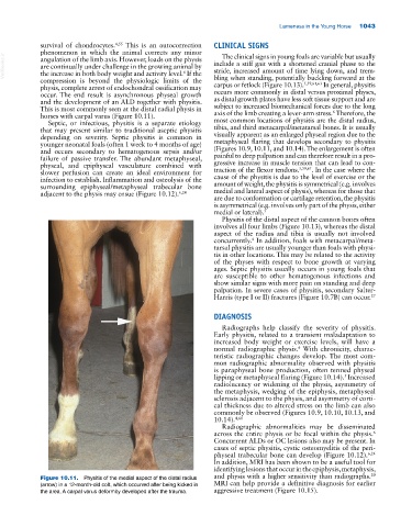
This is most commonly seen at the distal radial physis in subject to increased biomechanical forces due to the long
6
horses with carpal varus (Figure 10.11). axis of the limb creating a lever‐arm stress. Therefore, the
Septic, or infectious, physitis is a separate etiology most common locations of physitis are the distal radius,
that may present similar to traditional aseptic physitis tibia, and third metacarpal/metatarsal bones. It is usually
depending on severity. Septic physitis is common in visually apparent as an enlarged physeal region due to the
younger neonatal foals (often 1 week to 4 months of age) metaphyseal flaring that develops secondary to physitis
and occurs secondary to hematogenous sepsis and/or (Figures 10.9, 10.11, and 10.14). The enlargement is often
failure of passive transfer. The abundant metaphyseal, painful to deep palpation and can therefore result in a pro
physeal, and epiphyseal vasculature combined with gressive increase in muscle tension that can lead to con
5,59,61
slower perfusion can create an ideal environment for traction of the flexor tendons. . In the case where the
infection to establish. Inflammation and osteolysis of the cause of the physitis is due to the level of exercise or the
surrounding epiphyseal/metaphyseal trabecular bone amount of weight, the physitis is symmetrical (e.g. involves
adjacent to the physis may ensue (Figure 10.12). 6,24 medial and lateral aspect of physis), whereas for those that
are due to conformation or cartilage retention, the physitis
is asymmetrical (e.g. involves only part of the physis, either
medial or lateral). 5
Physitis of the distal aspect of the cannon bones often
involves all four limbs (Figure 10.13), whereas the distal
aspect of the radius and tibia is usually not involved
4
concurrently. In addition, foals with metacarpal/meta
tarsal physitis are usually younger than foals with physi
tis in other locations. This may be related to the activity
of the physes with respect to bone growth at varying
ages. Septic physitis usually occurs in young foals that
are susceptible to other hematogenous infections and
show similar signs with more pain on standing and deep
palpation. In severe cases of physitis, secondary Salter‐
Harris (type I or II) fractures (Figure 10.7B) can occur. 27
DIAGNOSIS
Radiographs help classify the severity of physitis.
Early physitis, related to a transient maladaptation to
increased body weight or exercise levels, will have a
normal radiographic physis. With chronicity, charac
6
teristic radiographic changes develop. The most com
mon radiographic abnormality observed with physitis
is paraphyseal bone production, often termed physeal
5
lipping or metaphyseal flaring (Figure 10.14). Increased
radiolucency or widening of the physis, asymmetry of
the metaphysis, wedging of the epiphysis, metaphyseal
sclerosis adjacent to the physis, and asymmetry of corti
cal thickness due to altered stress on the limb can also
commonly be observed (Figures 10.9, 10.10, 10.13, and
10.14). 4,61
Radiographic abnormalities may be disseminated
6
across the entire physis or be focal within the physis.
Concurrent ALDs or OC lesions also may be present. In
cases of septic physitis, cystic osteomyelitis of the peri
physeal trabecular bone can develop (Figure 10.12). 6,24
In addition, MRI has been shown to be a useful tool for
identifying lesions that occur in the epiphysis, metaphysis,
20
Figure 10.11. Physitis of the medial aspect of the distal radius and physis with a higher sensitivity than radiographs.
(arrow) in a 12‐month‐old colt, which occurred after being kicked in MRI can help provide a definitive diagnosis for earlier
the area. A carpal varus deformity developed after the trauma. aggressive treatment (Figure 10.15).