
Page 1079 - Adams and Stashak's Lameness in Horses, 7th Edition
P. 1079
VetBooks.ir
A B
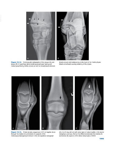
Figure 10.14. Craniocaudal radiographs of the carpus (A) and (black arrows) and radiolucency at the level of the medial physis
tarsus (B) of weanlings demonstrating paraphyseal lipping and (black arrowhead) causing widening of the physis.
metaphyseal flaring (white arrows) as well as metaphyseal sclerosis
A B C
Figure 10.15. Proton density sequences (1.5 T) of sagittal slices (B) of an 8‐day‐old colt with early signs of osteomyelitis in the lateral
through the medial (A) and lateral (C) aspects of the metaphysis (arrows). Notice the difference between the medial (A)
metacarpophalangeal joint shown in the dorsopalmar radiograph and lateral (B) aspects of the distal metaphysis of MCIII.
1045