
Page 1074 - Adams and Stashak's Lameness in Horses, 7th Edition
P. 1074
1040 Chapter 10
successful internal fixation with lag screws and casting articular shearing force or is secondary to infectious
31
has been described. Internal fixation (medial bone plate physitis. Open reduction and internal fixation are neces
VetBooks.ir mandatory for type II proximal tibial fractures. 42,51 opment of secondary osteoarthritis (OA).
sary to reconstruct the joint surface to prevent the devel
and/or screw and wire transphyseal bridge) is usually
Type III Type IV
Type III fracture is intra‐articular and extends from Type IV fractures are intra‐articular and extend from
the joint surface, through the epiphysis, into the deep the joint surface through the epiphysis, across the entire
zone of the growth plate and then along the physis to its thickness of the physeal plate, and through a portion
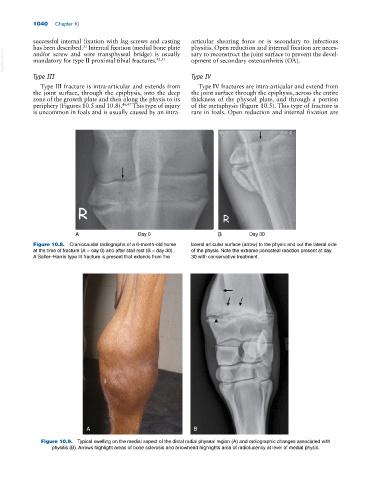
periphery (Figures 10.5 and 10.8). 46,47 This type of injury of the metaphysis (Figure 10.5). This type of fracture is
is uncommon in foals and is usually caused by an intra‐ rare in foals. Open reduction and internal fixation are
A Day 0 B Day 30
Figure 10.8. Craniocaudal radiographs of a 6‐month‐old horse lateral articular surface (arrow) to the physis and out the lateral side
at the time of fracture (A = day 0) and after stall rest (B = day 30). of the physis. Note the extreme periosteal reaction present at day
A Salter–Harris type III fracture is present that extends from the 30 with conservative treatment.
A B
Figure 10.9. Typical swelling on the medial aspect of the distal radial physeal region (A) and radiographic changes associated with
physitis (B). Arrows highlight areas of bone sclerosis and arrowhead highlights area of radiolucency at level of medial physis.