
Page 1072 - Adams and Stashak's Lameness in Horses, 7th Edition
P. 1072
1038 Chapter 10
VetBooks.ir
Type 1 Type 2 Type 3
Type
Type 4 Type 5 Type 6
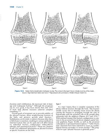
Figure 10.5. Salter–Harris classification of physeal injuries. The arrows in the type V injury indicate crushing of the physis.
48
Source: Reprinted from Salter and Harris. Reproduced with permission of Wolters Kluwer Health Inc.
duration until stabilization, the increased risk of dam Type I
age and inadequate healing. Although the prognosis In a type I injury, there is complete separation of the
51
can be very good, it has been shown that only 25%
of foals with physeal fractures achieved complete physis without fracture through the bone. This fracture
usually occurs due to shearing forces across the physis
soundness. 16,42
Overall goals of treatment are to provide stability of causing the fracture to propagate through the zone of
the physeal fracture, allow weight‐bearing function of hypertrophy with the germinal cells of the growth plate
39
the limb, and preserve growth plate function. remaining with the epiphysis (Figures 10.5 and 10.6).
51
With external trauma, this fracture configuration is com
Stabilization can be tricky to achieve because often there
is little bone to purchase, resulting in various methods of mon in the proximal femoral physis (slipped capital fem
oral epiphysis; Figure 10.6B), as well as the proximal
internal fixation that can be used and will differ based 4,55
on individual fracture configuration (Salter–Harris radial physis or proximal humeral physis, Salter‐Harris
type I fractures have been reported to occur as a patho
subtype) and anatomical location. Please see Chapters 27
33
4 and 5 for additional information on physeal fractures logical fracture secondary to severe physitis. The prog
27
nosis for type I physeal fractures is generally poor.
at specific locations on the limb.