
Page 339 - Adams and Stashak's Lameness in Horses, 7th Edition
P. 339
VetBooks.ir
A
B
C
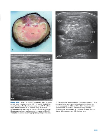
Figure 3.90. (A) In Z1A the SDFT is round but with a flat border (C) This ultrasound image is taken at the proximal aspect of Z1A to
dorsally where it is adjacent to the DDFT (A and B). The DDFT is correspond to the gross section slice plane that is close to the
rounded in shape. The ICL is located dorsal to the DDFT with the carpometacarpal joint. At this level, the ICL is the only structure
carpal sheath visualized as an anechoic triangular structure apparent dorsal to the DDFT. The median artery and large
between these two structures (B). The SL is located dorsal to the metacarpal vein are prominent on the medial aspect of the DDFT.
ICL but may only have a few fibers apparent in proximal Z1A (C). Source: US images courtesy of Dr. Caitlyn Horne.
The SL becomes more apparent, progressing distally in this zone.
305