
Page 341 - Adams and Stashak's Lameness in Horses, 7th Edition
P. 341
VetBooks.ir
B
A
C D
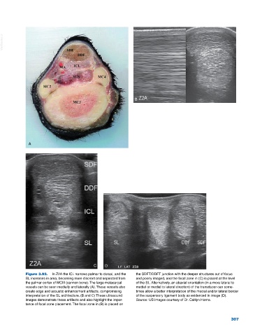
Figure 3.92. In Z2A the ICL narrows palmar to dorsal, and the the SDFT/DDFT junction with the deeper structures out of focus
SL increases in area, becoming more discreet and separated from and poorly imaged, and the focal zone in (C) is placed at the level
the palmar cortex of MCIII (cannon bone). The large metacarpal of the SL. Alternatively, an abaxial orientation (in a more lateral to
vessels can be seen medially and laterally (A). These vessels also medial or medial to lateral direction) of the transducer can some
create edge and acoustic enhancement artifacts, compromising times allow a better interpretation of the medial and/or lateral border
interpretation of the SL architecture. (B and C) These ultrasound of the suspensory ligament body as evidenced in image (D).
images demonstrate these artifacts and also highlight the impor Source: US images courtesy of Dr. Caitlyn Horne.
tance of focal zone placement. The focal zone in (B) is placed on
307