
Page 342 - Adams and Stashak's Lameness in Horses, 7th Edition
P. 342
308 Chapter 3
A B
VetBooks.ir
C
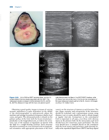
Figure 3.93. (A) In Z2B the SDFT becomes flatter, and the ICL cially demonstrates the fibers of the SDFT/DDFT interface, while
remains distinct from but closely associated with the DDFT. The (C) shows focal zone at the level of the SL and the convergence of
metacarpal vessels converge to midline between the ICL and the the large metacarpal vessels palmar to the SL. Source: US images
SL. (B) This ultrasound image with the focal zone placed superfi courtesy of Dr. Caitlyn Horne.
Obtaining a good‐quality images in tissues of varying zone(s) on the structure of interest at each location. The
acoustic impedances is quite dependent on the ability size, shape, margins, and echogenicity of the structures
of the ultrasonographer to appropriately adjust the should be evaluated, and a measurement system, using
machine and settings (transducer, frequency, depth, focal distance (cm) or zones, should be used to obtain images
zones, and gain). The ultrasound probe is placed on the at regular intervals. Comparison to the contralateral
palmar surface of the limb with the beam oriented per limb is important in all cases, even those with obvious
pendicular to the longitudinal axis, as well as the trans injury in the primary limb. In the metacarpus each level
verse axis of the tendons or ligaments of interest with should have two transverse scans performed. The first
the limb in a weight‐bearing position. Each structure scan should have a standoff placed on the transducer
should be examined on cross‐sectional and on longitudi with the focal zone(s) and transducer angle directed ini
nal orientation with appropriate placement of the focal tially at the superficial digital flexor (SDFT) and deep digital