
Page 343 - Adams and Stashak's Lameness in Horses, 7th Edition
P. 343
VetBooks.ir
B
A
C
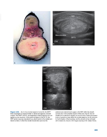
Figure 3.94. (A). In the proximal aspect of zone 3A, the SDFT predominant arterial blood supply to the DDF within the sheath
thins and begins to expand medial to lateral and appears crescent (arrows) (B). In the proximal aspect of this zone, the SL has not
shaped. The DDFT and ICL are beginning to blend together and can divided and continues to appear as one structure. There may appear
appear as one structure. In the proximal aspect of the DFTS, the to be a hypoechoic area within the central aspect of the SL just prior
DDF tendon receives mesotendinous attachments to the medial and to the formation of the branches, but this should not be confused
lateral borders of which the medial mesotendon provides the with a lesion (C). Source: US images courtesy of Dr. Caitlyn Horne.
309