
Page 345 - Adams and Stashak's Lameness in Horses, 7th Edition
P. 345
VetBooks.ir
B
A
C D
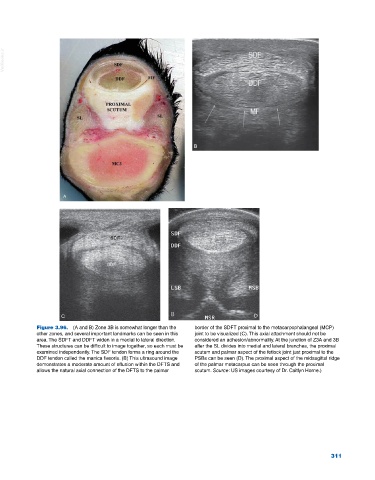
Figure 3.96. (A and B) Zone 3B is somewhat longer than the border of the SDFT proximal to the metacarpophalangeal (MCP)
other zones, and several important landmarks can be seen in this joint to be visualized (C). This axial attachment should not be
area. The SDFT and DDFT widen in a medial to lateral direction. considered an adhesion/abnormality. At the junction of Z3A and 3B
These structures can be difficult to image together, so each must be after the SL divides into medial and lateral branches, the proximal
examined independently. The SDF tendon forms a ring around the scutum and palmar aspect of the fetlock joint just proximal to the
DDF tendon called the manica flexoria. (B) This ultrasound image PSBs can be seen (D). The proximal aspect of the midsagittal ridge
demonstrates a moderate amount of effusion within the DFTS and of the palmar metacarpus can be seen through the proximal
allows the natural axial connection of the DFTS to the palmar scutum. Source: US images courtesy of Dr. Caitlyn Horne.)
311