
Page 349 - Adams and Stashak's Lameness in Horses, 7th Edition
P. 349
VetBooks.ir
A
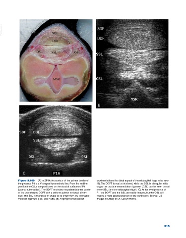
Figure 3.100. (A) In ZP1A the contour of the palmar border of proximad allows the distal aspect of the midsagittal ridge to be seen
the proximal P1 is a V‐shaped hyperechoic line. From the midline (B). The DDFT is oval at this level, while the SSL is triangular at its
position the OSLs are positioned on the abaxial surfaces of P1 origin; the cruciate sesamoidean ligament (CSL) can be seen dorsal
(palmar tuberosities). The SDFT encircles the palmar/plantar border to the SSL (and the midsagittal ridge). (C) At the level proximal of
of the oval‐shaped DDFT with a uniform palmar to dorsal dimen P1, the DDFT and the SSL are easily imaged, but the OSL will
sion. The SSL is triangular in shape at its origin from the intersesa require a more abaxial position of the transducer. Source: US
moidean ligament (ISL) and PSBs. (B) Angling the transducer images courtesy of Dr. Caitlyn Horne.
315