
Page 351 - Adams and Stashak's Lameness in Horses, 7th Edition
P. 351
VetBooks.ir
B
A
C
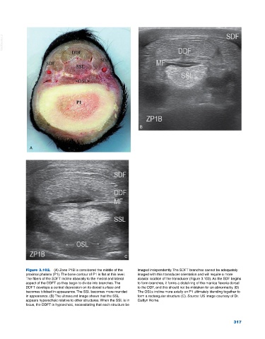
Figure 3.102. (A) Zone P1B is considered the middle of the imaged independently. The SDFT branches cannot be adequately
proximal phalanx (P1). The bone contour of P1 is flat at this level. imaged with this transducer orientation and will require a more
The fibers of the SDFT incline abaxially to the medial and lateral abaxial location of the transducer (Figure 3.103). As the SDF begins
aspect of the DDFT as they begin to divide into branches. The to form branches, it forms a distal ring of the manica flexoria dorsal
DDFT develops a central depression on its dorsal surface and to the DDF, and this should not be mistaken for an abnormality. (B)
becomes bilobed in appearance. The SSL becomes more rounded The OSLs incline more axially on P1 ultimately blending together to
in appearance. (B) The ultrasound image shows that the SSL form a rectangular structure (C). Source: US image courtesy of Dr.
appears hyperechoic relative to other structures. When the SSL is in Caitlyn Horne.
focus, the DDFT is hypoechoic, necessitating that each structure be
317