
Page 352 - Adams and Stashak's Lameness in Horses, 7th Edition
P. 352
318 Chapter 3
VetBooks.ir
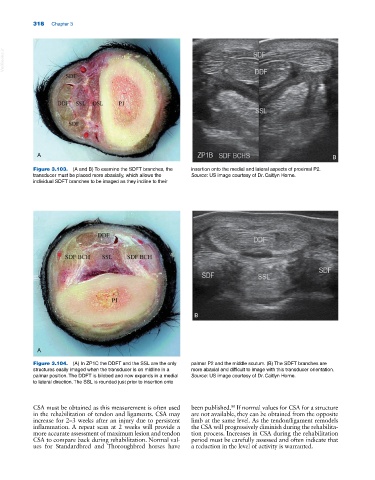
A B
Figure 3.103. (A and B) To examine the SDFT branches, the insertion onto the medial and lateral aspects of proximal P2.
transducer must be placed more abaxially, which allows the Source: US image courtesy of Dr. Caitlyn Horne.
individual SDFT branches to be imaged as they incline to their
B
A
Figure 3.104. (A) In ZP1C the DDFT and the SSL are the only palmar P2 and the middle scutum. (B) The SDFT branches are
structures easily imaged when the transducer is on midline in a more abaxial and difficult to image with this transducer orientation.
palmar position. The DDFT is bilobed and now expands in a medial Source: US image courtesy of Dr. Caitlyn Horne.
to lateral direction. The SSL is rounded just prior to insertion onto
CSA must be obtained as this measurement is often used been published. If normal values for CSA for a structure
80
in the rehabilitation of tendon and ligaments. CSA may are not available, they can be obtained from the opposite
increase for 2–3 weeks after an injury due to persistent limb at the same level. As the tendon/ligament remodels
inflammation. A repeat scan at 2 weeks will provide a the CSA will progressively diminish during the rehabilita
more accurate assessment of maximum lesion and tendon tion process. Increases in CSA during the rehabilitation
CSA to compare back during rehabilitation. Normal val period must be carefully assessed and often indicate that
ues for Standardbred and Thoroughbred horses have a reduction in the level of activity is warranted.