
Page 356 - Adams and Stashak's Lameness in Horses, 7th Edition
P. 356
322 Chapter 3
occurs when the ultrasound beam is not at 90° to the
fibers of the target structure that reflect the returning
VetBooks.ir echoic area that mimics a lesion(s) within the targeted
echoes away from the transducer. This creates a hypo
structure. Improper gain and power settings and inap
propriate focal zone position can also lead to subopti
mal images. Near‐gain and power settings that are set
too high reduce the ability to differentiate the tissues.
Gain settings should be adjusted to produce a uniform
gray scale across the entire image. Focal zones are
variable in number and position and should be adjusted
to the level of the specific structure(s) to optimize the
image quality.
Artifacts that are created by sound–tissue interac
tions include acoustic enhancement, refractive scattering
(edge artifacts), reverberation, and acoustic shadowing.
Acoustic enhancement occurs when sound passes
through a fluid structure. Fluid attenuates sound less
than soft tissue. Enhancement results from relatively
increased amplitude of deeper echoes by an overlying RH PSD
structure of low attenuation. This creates the appear Z1A
ance of increased echogenicity of the tissue deep to the 24 CM DPOH
fluid‐filled structure (far‐field enhancement). The most
common structures that can cause enhancement arti
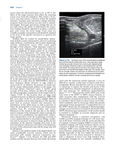
facts in the musculoskeletal examinations are veins and Figure 3.110. Transverse scan of the proximal plantar metatarsal
arteries. Refractive artifacts are generated from the edge area with the inherent artifacts that occur in this area due to edge
of a curved surface and are created because part of the shadowing, acoustic enhancement, and acoustic shadowing that
sound beam is refracted off the curved structure and occur because of the bony anatomy and overlying SDF and DDF
does not return to the transducer. Shadow artifacts are and vessels. This lesion was found to be a false positive when the
displayed distal the lateral margins of curved or cystic horse had a normal MRI examination of this area. The operator must
find an acoustic window that minimizes the interference of the splint
structures. Edge artifacts are consistent and particularly bones as well as prevents or limits the development of refraction and
troublesome when examining specific anatomical loca enhancement artifacts from the overlying tendons and vessels.
tions. The SDF, DDF, and ICL are superimposed over the
SL in the metacarpus and metatarsus, and the size dif
ferential (SL is wider than the SDF/DDF/ICL) between canal with the restricting annular ligament, it may be
these structures allows refractive or edge artifact to difficult to visualize certain types of injuries with ultra
occur in the SL. In addition, the presence of vessels over sonography. Recent introduction and use of tenoscopy
the SL creates enhancement artifacts in the image. The or endoscopy to explore the sheaths and bursae of the
proximal plantar metatarsal region is another good equine limb have allowed comparison of ultrasonogra
example of how these artifacts can affect the quality of phy and tenoscopy for identifying tendon/ligament
the ultrasonographic examination of the origin of the SL injury. 37,75,76,92,104 Tenosynovitis of the DFTS may result
(Figure 3.110). At this location there are large oval/ from tears in the DDFT or SDFT, manica flexoria, or
rounded structures of different echogenicity (SDF, DDF, other structures that communicate with the synovial
ICL, and origin of the SL) all with a slightly different fluid and thus can generate an intense synovial response
fiber orientation and acoustic impedances. The operator in a sheath or bursa. However, these lesions can be iden
must find an acoustic window that minimizes the tified confidently only by tenoscopy, which also permits
interference of the splint bones as well as prevents or appropriate lesion management. Longitudinal tears of
92
limits the development of refraction and enhancement the DDFT should also be suspected if ultrasonographic
artifacts from the overlying tendons and vessels. changes are present lateral or medial to the border of the
Dilatation of the vessels in this area due to the inflam DDFT, but tenoscopic examination of the tendon sheath
matory response of the damaged SL makes scanning this is essential to establish an accurate diagnosis and an
area even more problematic and significantly compro effective treatment. 104
mises interpretation of the SL morphology. Scanning at Ultrasound remains a practical, inexpensive, and
a more plantaromedial to dorsolateral oblique orienta readily accessible imaging technique for soft tissue inju
tion may be necessary to improve image quality, but the ries of the horse. However, with the recent introduction
experience of the operator scanning areas such as the of magnetic resonance imaging (MRI) to equine muscu
proximal plantar metatarsus will improve the diagnostic loskeletal imaging, soft tissue and bone injuries can be
accuracy of the ultrasound exam, but the image may still evaluated in detail not capable with any other imaging
be suboptimal. technique. MRI is now considered the gold standard to
Injuries to the tendons inside of the digital sheath can assess lameness originating from the carpus and tarsus
cause considerable synovial reaction, which may be distally (especially of the foot within the horny hoof
associated with annular ligament constriction syn capsule), but it is not always practical or necessary to
drome 28,33 (Figure 3.111). Because the SDF and DDF make a diagnosis. Lesion(s) seen with standard ultra
tendons and the macular flexoria exist inside the fetlock sonographic imaging are not typically candidates for