
Page 354 - Adams and Stashak's Lameness in Horses, 7th Edition
P. 354
VetBooks.ir
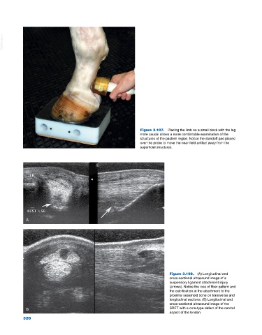
Figure 3.107. Placing the limb on a small block with the leg
more caudal allows a more comfortable examination of the
structures of the pastern region. Notice the standoff pad placed
over the probe to move the near‐field artifact away from the
superficial structures.
A
Figure 3.108. (A) Longitudinal and
cross‐sectional ultrasound image of a
suspensory ligament attachment injury
(arrows). Notice the loss of fiber pattern and
the calcification at the attachment to the
proximal sesamoid bone on transverse and
longitudinal sections. (B) Longitudinal and
cross‐sectional ultrasound image of the
B SDFT with a core‐type defect of the central
aspect of the tendon.
320
320