
Page 355 - Adams and Stashak's Lameness in Horses, 7th Edition
P. 355
Diagnostic Imaging 321
denser the structure is, the more echoes it returns, and
the whiter the structure appears.
VetBooks.ir don and ligamentous structures. The fibers of tendinous
Fibers have a parallel alignment in most normal ten
tissue are more uniformly distributed, while the fibers in
ligamentous tissue are more multidirectional. This par
allel fiber bundle alignment is best assessed on longitu
dinal images. Injury to and inflammation of tendons and
ligaments can disrupt fiber bundle alignment. Subtle
changes in fiber alignment are best seen on the longitu
dinal plane images. More severe fiber bundle alignment
changes can begin to be appreciated on transverse
images. The longitudinal image must be obtained
through the affected tissue as seen on transverse images.
Damage seen on cross section should be confirmed on
longitudinal orientation. Fiber disruption seen as echo
lucent areas surrounding the fibers is compatible with
hemorrhage and edema seen with acute injuries.
Nonparallel or random fiber alignment without echolu
cent fluid content is compatible with chronic injury.
Tendon injury can be focal or generalized such that the
distribution of fiber damage can be quite variable.
Fibroblasts migrate into the damaged area and begin to
deposit collagen and form granulation tissue. This col
lagen is laid down randomly, and cross‐links are pro
duced between the fibers. This random disorganized
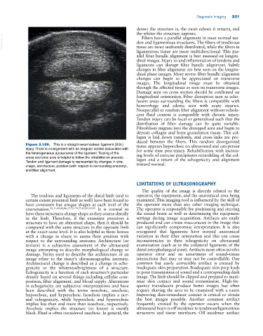
Figure 3.109. This is a straight sesamoidean ligament (SSL) tissue appears hypoechoic on ultrasound and can persist
injury. There is enlargement with an irregular outline associated with for some time post‐injury. Rehabilitation with increas
the heterogeneous appearance of the ligament. Tracing of the ing levels of exercise precipitates remodeling of the col
cross‐sectional area is helpful to follow the rehabilitation process. lagen and a return of the echogenicity and alignment
Tendon and ligament damage is represented by changes in size, toward normal.
shape, architecture, position (with respect to surrounding anatomy),
and fiber alignment.
LIMITATIONS OF ULTRASONOGRAPHY
The quality of the image is directly related to the
The tendons and ligaments of the distal limb (and to operator, the equipment, and the anatomical area being
certain extent proximal limb as well) have been found to examined. This imaging tool is influenced by the skill of
have consistent but unique shapes at each level of the the operator more than any other imaging technique.
examination. 22,31,40,41,46,57,72–74,77,82,85,90,99 It is normal to The operator is responsible for positioning and steering
have these structures change shape as they course distally the sound beam as well as determining the equipment
in the limb. Therefore, if the examiner perceives a settings during image acquisition. Artifacts are easily
structure to have an abnormal shape, then it should be produced and can create inaccuracies in the image that
compared with the same structure in the opposite limb can significantly compromise interpretation. It is also
at the exact same level. It is also helpful in those horses recognized that ligaments have normal anatomical
with a change in shape to assess their position with variation in their fiber orientation and this can cause
respect to the surrounding anatomy. Architecture (or inconsistencies in their echogenicity on ultrasound
texture) is a subjective assessment of the ultrasound examination (such as in the collateral ligaments of the
image attempting to describe morphological change or distal interphalangeal joint). Artifacts most often involve
damage. Terms used to describe the architecture of an operator error and an assortment of sound–tissue
image relate to the tissue’s ultrasonographic intensity. interactions that may or may not be controllable. One
Architectural change is described as a change in echo common but easily correctible artifact is created by
genicity or the whiteness/brightness of a structure. inadequate skin preparation. Inadequate skin prep leads
Echogenicity is a function of each structure’s particular to poor transmission of sound and a corresponding dark
density based on several things including cellular com image. The limb should be clipped and prepped to maxi
position, fiber alignment, and blood supply. Alterations mize skin contact and sound transmission. High‐fre
in echogenicity are subjective interpretations and have quency transducers produce better images but often
been described with the terms isoechoic, anechoic, require shaving the area to be examined with a razor.
hypoechoic, and hyperechoic. Isoechoic implies a nor Improving skin–transducer contact is critical to obtain
mal echogenicity, while hypoechoic and hyperechoic the best images possible. Another common artifact
implies less than and more than isoechoic, respectively. frequently created by the operator occurs when the
Anechoic implies the structure (or lesion) is mostly ultrasound beam is off incidence to tendinous/ligamentous
black. Fluid is often considered anechoic. In general, the structures and tissue interfaces. Off incidence artifact