
Page 360 - Adams and Stashak's Lameness in Horses, 7th Edition
P. 360
326 Chapter 3
ULTRASONOGRAPHIC APPEARANCE extended and relaxed, whereas while the limb is flexed,
the short collateral ligaments are tensed only when the
OF PERIARTICULAR STRUCTURES
VetBooks.ir Collateral Ligaments tarsus is flexed. Therefore, the short collateral liga
99
ments should be examined while the leg is flexed. (See
tarsus and tibia in Chapter 5 for more information.)
The joints of the appendicular skeleton in the horse
are designed to work predominantly in the sagittal plane
with flexion and extension as the primary range of Tendons and Ligaments
motion. A major component of the stability of a joint is
provided by the periarticular soft tissues but in particu As discussed previously tendons and ligaments
lar the collateral ligaments. The collateral ligaments are appear as moderately echogenic structures with relatively
designed to impart stability to the joint throughout its well‐defined margins, and injury is recognized with
entire range of motion. This unique task is accomplished ultrasound by changes in size, shape, architecture, posi
either with paired structures (i.e. multiple bundles of tion, and fiber alignment. Ultrasonographic examina
the fetlock, tarsal joints) or by unpaired structures (i.e. tion of the tendons at the level of the joint is no different
single bundle of coffin joint). Knowing the anatomic except from a couple of perspectives. First, the examiner
arrangement of the collateral ligaments of each specific must pay particular attention to the course of the tendon
joint becomes extremely important as the ultrasono or ligament as it courses over a joint because there can
graphic appearance of these ligaments is determined by be quite remarkable changes in direction. This becomes
the fiber orientation. Most of the collateral ligaments important in that the transducer must constantly be
have a uniform fascicular orientation and, therefore, a adjusted to maintain a perpendicular orientation to the
homogeneous ultrasonographic appearance. Some have structure to prevent beam angle artifact. Second, while
a mixed fascicular arrangement, which gives them a het injury is recognized as in other areas, the periarticular
erogeneous appearance. The lateral collateral ligaments portions of tendons have sheaths (and occasionally
of the stifle, hock, and elbow and the collateral liga bursae) associated with them. Injury to these tendons/
ments of the fetlock and carpus have spiral or crossed ligaments may also manifest as effusion within the
23
fibers. This mixed arrangement of fascicles within the sheaths or bursae. To confuse the matter even more, the
CLs is one of the arrangements that allow the collateral effusion may occur without structural damage to the
ligaments to function in both extension and flexion tendon or ligament. Therefore, effusion of the sheaths and
(Figure 3.112). In the tarsocrural joint, the superficial bursae associated with these structures requires careful
collateral ligaments are under tension while the limb is evaluation to define the cause and to differentiate the
fluid accumulation as separate from joint effusion.
A
B
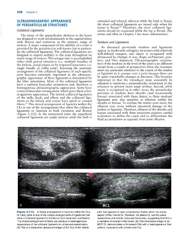
Figure 3.112. A mixed arrangement of fascicles within the CLs joint that appears to have a hypoechoic lesion within the dorsal
of many joints is one of the unique arrangements of ligaments that aspect of the medial CL. However, the lateral CL has the same
allow a collateral ligament to function in both extension and flexion. appearance and similar cross‐sectional area, suggesting that this is
This mixed arrangement of fibers can produce a heterogeneous a variation of normal in this horse. (B) This transverse image of an
appearance of the collateral ligament on ultrasound examination. MCP CL demonstrates a thickened CSA with a heterogeneous fiber
(A) This is a transverse ultrasound image of the CLs of the fetlock pattern, consistent with chronic desmitis.