
Page 364 - Adams and Stashak's Lameness in Horses, 7th Edition
P. 364
330 Chapter 3
VetBooks.ir
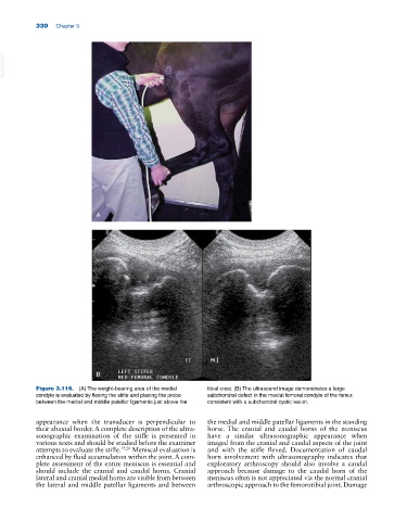
A
B
Figure 3.116. (A) The weight‐bearing area of the medial tibial crest. (B) The ultrasound image demonstrates a large
condyle is evaluated by flexing the stifle and placing the probe subchondral defect in the medial femoral condyle of the femur,
between the medial and middle patellar ligaments just above the consistent with a subchondral cystic lesion.
appearance when the transducer is perpendicular to the medial and middle patellar ligaments in the standing
their abaxial border. A complete description of the ultra horse. The cranial and caudal horns of the meniscus
sonographic examination of the stifle is presented in have a similar ultrasonographic appearance when
various texts and should be studied before the examiner imaged from the cranial and caudal aspects of the joint
attempts to evaluate the stifle. 17,20 Meniscal evaluation is and with the stifle flexed. Documentation of caudal
enhanced by fluid accumulation within the joint. A com horn involvement with ultrasonography indicates that
plete assessment of the entire meniscus is essential and exploratory arthroscopy should also involve a caudal
should include the cranial and caudal horns. Cranial approach because damage to the caudal horn of the
lateral and cranial medial horns are visible from between meniscus often is not appreciated via the normal cranial
the lateral and middle patellar ligaments and between arthroscopic approach to the femorotibial joint. Damage