
Page 367 - Adams and Stashak's Lameness in Horses, 7th Edition
P. 367
Diagnostic Imaging 333
VetBooks.ir
B
A
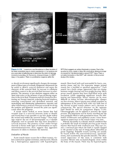
Figure 3.119. Lacerations over the extensor or flexor tendons of DFTS that suggests an active inflammatory process. Due to the
the distal extremities require careful examination of the tendons and penetrating injury, sepsis is likely. (B) Longitudinal ultrasound image
any associated sheaths/bursae to determine the extent of damage of a laceration that demonstrates a severe DDFT injury. There is
incurred at wounding. (A) Transverse ultrasound image of a DDFT complete transaction of the DDFT with retraction of the proximal
injury in the pastern. There is a proliferative response within the stump of the tendon.
or sheath involvement significantly changes the manage muscle fibers laced with and surrounded by fascia, con
ment of these types of wounds. Diagnostic ultrasound can nective tissue, and fat. On transverse images normal
be useful to identify synovial distension and assess the muscle has a marbled or speckled appearance. Each
81
character of the synovial fluid. An increase in cellularity muscle has a fairly unique appearance that can change
and fibrin content in the synovial fluid increases its echo between weight‐bearing and non‐weight‐bearing. More
genicity. The presence of gas shadows suggests either an severe muscle injuries may have fluid‐filled areas with
open joint space or the presence of gas‐producing organ loculation visible suggesting significant muscle fiber
isms in the joint fluid. Treatment must be directed at elim tearing and hemorrhage. Minor injuries can be more
inating any foreign material, reducing bacterial numbers, difficult to define because ultrasonographic changes
removing contaminated and devitalized material, and are less obvious. Minor injuries may simply manifest an
neutralizing and eliminating inflammatory enzymes and enlargement of the muscle belly with very little loss of
other inflammatory products. Puncture wounds involving the normal striated muscle pattern and a decrease in
the tendons and ligaments around the joint can signifi echogenicity. The paired structure on the opposite
cantly affect the prognosis. limb should be used for comparison (Figure 3.120).
A recent report looked at seven horses that had Ultrasonography can be utilized to monitor the repair
wounds close to a synovial structure (joint or sheath) process. The hematoma will be slowly resorbed and the
and found that it was possible to see hair shafts within area gradually filled in with granulation tissue. The infil
65
the wound and within the synovial lining. These find tration of fibrocytes and capillaries create a more heter
ings support the use of ultrasonography as a valuable ogeneous appearance to the muscle injury over time.
diagnostic tool in wounds that may have breached a Fibrotic myopathy is a chronic muscle condition that
synovial structure. The presence of hair and/or foreign is probably the result of multiple previous acute muscle
material in horses with signs of inflammatory or septic injuries that healed with exuberant scar. This scar cre
synovitis/tenosynovitis often suggests that aggressive ates a restricted gait where the limb is rapidly slapped
measures be taken to eliminate the material. to the ground at the end of swing phase (described as
goose stepping). Palpable muscle scarring can usually
Evaluation of Muscle be found within the semimembranosus, semitendi
nosus, and occasionally the gracilis muscles. The ultra
Acute muscle injury occurs due to blunt trauma, vio sound appearance of this mass is consistent with
lent contraction against resistance, and myositis. Muscle excessive fibrosis seen as hyperechogenic areas within
has a heterogeneous appearance with hypoechogenic the body of the muscle. There may be hyperechogenic