
Page 369 - Adams and Stashak's Lameness in Horses, 7th Edition
P. 369
Diagnostic Imaging 335
NEW DIRECTIONS IN THE USE OF ULTRASOUND a sterile ultrasound covers). As a means to improve nee
dle placement and minimize damage to vessels in the
VetBooks.ir Diagnostic ultrasound can enhance the accurate determine a path for the needle free of vessels that may
Ultrasound‐Guided Injections
area, a Doppler examination (color or power) can help
not be obvious on gray scale. As will be discussed in a
placement of a needle into a specific site. Real‐time nee
dle tracking allows the needle to be visualized entering a later section on Doppler examination of soft tissue
target area (Figure 3.122). For example, the correct injury, power Doppler (PD) can provide some insight of
placement of diagnostic analgesia will help improve the where within a structure an injection should be directed
accuracy of diagnosing the cause of lameness, and plac to have the most advantageous effect.
ing medication or regenerative therapy (platelet‐rich Sterile technique requires a large area of skin prep
plasma [PRP], stem cells, and interleukin receptor antag with chlorhexidine detergent and alcohol rinse. It is
onist protein [IRAP]) directly into a lesion will enhance advisable to disinfect the probe and cable with a disin
the successful treatment of that injury. In veterinary fectant before placement of a sterile probe cover or
medicine there are several reports evaluating the use of surgical glove. The examiner should view the area
diagnostic ultrasound to inject the navicular bursa, cer between the probe and the structure to be injected.
vical articular facets, scapulohumeral joint, coxofemoral Previewing the area and knowing the pertinent local
joint, and the sacroiliac joint. 5,10,14,16,60,93 Competency in anatomy are critical to avoid penetration of key struc
ultrasound‐guided injection technique begins with a tures (vessels and nerves). It is best to mapped out the
comprehensive knowledge of the anatomy of the area approach that will be taken to drive the needle to the
both grossly and as seen with ultrasound. A reasonable target. A decision should be made about utilizing short
amount of skill is required to handle a transducer axis or long axis visualization of the needle. The exam
focused on the area of interest while driving a needle iner should use the nondominant hand to manipulate
within the ultrasound beam. Depth of the structure to be the probe and scan the target keeping 2–3 fingers of
injected should determine the angle the needle must take the probe hand in contact with the patient’s skin. Most
to hit the structure. To increase the degree of difficulty, often the needle should cross the longitudinal plane of
ultrasound‐guided injections are frequently done while the sound beam, and the examiner then must make
maintaining sterile technique (the probe is covered with short axis slides of the probe to keep the needle in
view. Placing the needle in the plane of the sound beam
(long axis) is preferred during most procedures because
the needle tip and shaft are visualized throughout the
entire procedure. Some ultrasound probes have a nee
dle guide that can be attached directly to the transducer
for accurate placement of the needle. Alternatively,
and probably more commonly, the needle may be
placed freehand. Either way, the needle should be
visualized throughout the procedure. The shortest
pathway to the target should be selected avoiding
regional neurovascular structures. Deeper injections
require more skill as tracking the needle can be more
challenging and requires better technique. Some alter
native techniques to improve ultrasound‐guided injec
tions include using beam steer, scoring the needle with
scalpel, using reflective facet tip needles, and placing
the bevel of the needle up. Another option though not
preferable is to use a blinded technique whereby a nee
dle is placed into the area of the synovial structures. If
no fluid is retrieved, then the needle position is evalu
ated with ultrasound to confirm the correct placement.
If it appears that the needle is position correctly, then
fluid can be injected to confirm the correct position
within the joint or any indication that repositioning
the needle is necessary.
MRI/Ultrasound Fusion: Navigational Ultrasound
MRI/ultrasound fusion imaging or navigational
ultrasound imaging is a novel imaging technique devel
oped to combine cross‐sectional imaging (CT or MRI)
with real‐time ultrasound. This technique has been
developed and utilized in the human medicine to sample
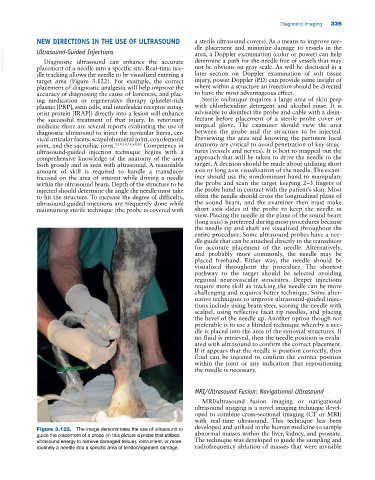
Figure 3.122. The image demonstrates the use of ultrasound to
guide the placement of a probe (in this picture a probe that utilizes abnormal masses within the liver, kidney, and prostate.
ultrasound energy to remove damaged tissue), instrument, or more The technique was developed to guide the sampling and
routinely a needle into a specific area of tendon/ligament damage. radiofrequency ablation of masses that were invisible