
Page 365 - Adams and Stashak's Lameness in Horses, 7th Edition
P. 365
Diagnostic Imaging 331
VetBooks.ir
B
A
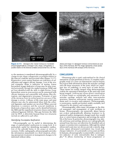
Figure 3.117. Damage to the medial meniscus is manifested ultrasound image of a damaged meniscus demonstrates an axial
ultrasonographically by a change in size, shape, echogenicity, or injury of the meniscus. (B) This image represents a more severe
position relative to the femoral condyles and proximal tibia. (A) This injury of the meniscus with prolapse of the meniscus.
to the meniscus is manifested ultrasonographically by a CONCLUSIONS
change in size, shape, echogenicity, or position relative to
the femoral condyles and proximal tibia (Figure 3.117). Ultrasonography is vastly underutilized in the clinical
Hyperechoic areas casting shadows are indicative of cal assessment of joint problems in horses. A complete radio
cification and suggest chronic damage. Loss of the nor graphic study of a joint can demonstrate osseous lesions,
mal triangular shape is indicative of tearing. Linear but it gives little information about the articular cartilage
hypoechoic images indicative of tears may be seen cross and soft tissue structures of the joint, which are impor
ing horizontally through the medial meniscus (MM) and tant sites of pathology in most types of joint disease.
are best identified with the stifle in slight flexion. Large These tissues are readily imaged using ultrasonography
hypoechoic areas in cross section can be associated with (within the anatomical limitations of the particular joint
chronic lameness and compatible with edema, fiber dis being imaged). Arthroscopy allows direct visualization
ruption, and degenerative processes. Extrusion of part of lesions found on radiography and assessment of the
of the meniscus from its normal position can occur if the articular cartilage surfaces and other intra‐articular
damage is severe. Collapse of the joint space and joint structures. However, arthroscopy requires general anes
effusion may also be appreciated when both the collat thesia and it is invasive and expensive. Ultrasonography
eral ligaments and menisci are involved. When synovial is noninvasive, rapidly performed, widely available, well
distension is extensive and the synovial membrane is tolerated by the patient, and inexpensive.
thickened, a hypoechoic space can be identified between Ultrasonography offers many advantages in the clini
the MM and the superficial structures (fascia and medial cal assessment and management of joint problems in
collateral ligament [MCL]). This gap between the MM horses. Ultrasonography can potentially detect lesions
and the MCL is abnormal, because these two structures not evident radiographically allowing treatment to be
normal are adhered to each other. instituted and/or management changes made that would
slow or arrest lesion progression and prolong the useful
Identifying Incomplete Ossification life of the horse. In particular, ultrasonography may iden
tify soft tissue and cartilage defects over radiographically
Ultrasonography can be useful in determining the normal bone that may not otherwise be identified only at
stage of endochondral ossification in young foals. It is surgery or postmortem examination. Although the initial
particularly useful for identifying incomplete ossifica ultrasound exam may not always provide a specific diag
tion of the cuboidal bones in the carpus or tarsus of nosis, repeat ultrasound examination may reveal early
neonates. Unlike radiography, ultrasonography provides changes indicative of a potentially serious joint problem.
the information immediately in a field situation, allow In humans, ultrasonography is purportedly a more sensi
ing appropriate treatment or management changes to be tive indicator of early osteoarthritis than is radiogra
instituted without delay. phy. It allows monitoring of lesion progression or
58