
Page 361 - Adams and Stashak's Lameness in Horses, 7th Edition
P. 361
Diagnostic Imaging 327
ULTRASONIC APPEARANCE OF THE JOINT increases the cellularity and fibrin deposition within the
joint and appears as echogenic fluid. Ultrasonography can
VetBooks.ir The joint capsule is primarily connective tissue with fluid accumulation. Utilization of this technique, particu
Joint Capsule, Synovium, and Synovial Fluid
aid in aspiration of synovial fluid by identifying areas of
larly in the deeper joints such as the hip and the shoulder,
low cell density. The joint capsule is continuous with the
periosteum or perichondrium, but it does not insert can minimize trauma to the joint and reduce the potential
directly at the perimeter of the articular cartilage for blood contamination of the fluid sample.
(Figure 3.113). There tends to be redundancy of the joint
capsule in the high‐motion joints, such as the fetlock, car
pus, tarsus, and stifle. In fact, this redundancy can cause
relaxation artifacts if the redundant aspect of the joint is
examined in the weight‐bearing position rather than dur
ing flexion. Inflammatory conditions within the joint can
cause capsular changes that include thickening (initially
due to hemorrhage and edema, and later fibrosis), calcifi
cation, and insertional capsulopathies (enthesophytes).
Osteoarthritis in high‐motion joints typically involves
periarticular changes that begin with congestion and
thickening of the joint capsule. The synovial membrane
becomes hyperplastic, and in more chronic cases, synovi
ocyte metaplasia may lead to the formation of synovial
chondromas, which are seen as nodules of cartilage. These
nodules may undergo endochondral ossification, result
ing in ovoid radiodense bodies within the joint capsule.
Synovitis is an early response to joint injury and usu
ally progresses to capsulitis; however, thickening of the
thin synovial membrane is difficult to appreciate ultra
sonographically. Synovial effusion is helpful in assessing
synovial membrane proliferation and thickening. In
cases of severe synovitis, synovial fluid that is fibrinous,
cellular, or hemorrhagic can cloud evaluation of the
joint capsule and synovium.
There is usually minimal synovial fluid in a normal
joint, so the joint capsule and synovium are normally in
close apposition with the articular cartilage. Synovial fluid
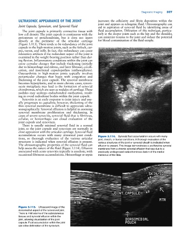
accumulation occurs with many abnormal joint condi Figure 3.114. Synovial fluid accumulation occurs with many
tions, so thorough evaluation of the various articular joint, sheath, or bursal conditions. A thorough evaluation of the
structures is indicated when synovial effusion is present. various structures of the joint or synovial sheath is indicated when
The ultrasonographic properties of the synovial fluid can effusion is present. This image demonstrates a proliferative synovial
help assess the nature of the fluid (Figure 3.114). Effusion membrane from a chronic synovial effusion that was due to a
associated with acute synovitis typically is anechoic, with previously undiagnosed osteochondrosis lesion of the medial
occasional fibrinous accumulations. Hemorrhage or sepsis malleolus of the tibia.
Figure 3.113. Ultrasound image of the
dorsomedial aspect of the tarsocrural joint.
There is mild edema of the subcutaneous
tissues and synovial effusion within the
joint, allowing visualization of the joint
capsule. Fluid accumulation within the joint
can allow delineation of the synovium.