
Page 363 - Adams and Stashak's Lameness in Horses, 7th Edition
P. 363
Diagnostic Imaging 329
VetBooks.ir
A
B
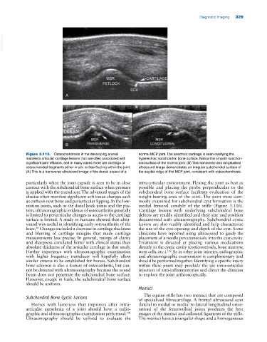
Figure 3.115. Osteochondrosis in the developing animal normal MCP joint. The anechoic cartilage is seen overlying the
manifests articular cartilage lesions that are often associated with hyperechoic subchondral bone surface. Notice the smooth subchon
significant joint effusion, and in many cases there are cartilage or dral surface of the normal joint. (B) This transverse and longitudinal
osteochondral fragments either in situ or free‐floating within the joint. ultrasound image demonstrates an irregular subchondral surface of
(A) This is a transverse ultrasound image of the dorsal aspect of a the sagittal ridge of this MCP joint, consistent with osteochondrosis.
particularly when the joint capsule is seen to be in close intra‐articular environment. Flexing the joint as best as
contact with the subchondral bone surface when pressure possible and placing the probe perpendicular to the
is applied with the transducer. The advanced stages of the subchondral bone surface facilitate evaluation of the
disease often manifest significant soft tissue changes such weight‐bearing area of the joint. The joint most com
as enthesis new bone and periarticular lipping. In the low‐ monly examined for subchondral cyst formation is the
motion joints, such as the distal hock joints and the pas medial femoral condyle of the stifle (Figure 3.116).
tern, ultrasonographic evidence of osteoarthritis generally Cartilage lesions with underlying subchondral bone
is limited to periarticular changes as access to the cartilage defects are readily identified and their size and position
surface is limited. A study in humans showed that ultra documented with ultrasonography. Subchondral cystic
sound was useful in identifying early osteoarthritis of the lesions are also readily identified and help characterize
knee. Changes included a decrease in cartilage thickness the size of the cyst opening and depth of the cyst. Some
58
and blurring of cartilage margins that made cartilage clinicians have reported using ultrasound to guide the
measurements less precise. In general, ratings of clarity placement of a needle percutaneously into the cyst cavity.
and sharpness correlated better with clinical status than Treatment is directed at placing various medications
absolute thickness of the articular cartilage in that study. directly in the cystic cavity (corticosteroids, bone marrow,
Further experience with ultrasonographic examination stem cells, etc.). As in other joint injuries, radiographic
102
with higher frequency transducer will hopefully allow and ultrasonographic examination is complementary and
similar criteria to be established for horses. Subchondral should be performed together. Identifying a specific injury
bone sclerosis is also a feature of osteoarthritis, but can within these joints may preclude the use intra‐articular
not be detected with ultrasonography because the sound injection of anti‐inflammatories and direct the clinician
beam does not penetrate the subchondral bone surface. to explore the joint arthroscopically.
However, except in foals, the subchondral bone surface
should be uniform.
Menisci
Subchondral Bone Cystic Lesions The equine stifle has two menisci that are composed
of specialized fibrocartilage. A frontal ultrasound scan
Horses with lameness that improves after intra‐ (lateral to medial or medial to lateral longitudinal orien
articular anesthesia of a joint should have a radio tation) of the femorotibial joints produces the best
graphic and ultrasonographic examination performed. images of the menisci and collateral ligaments of the stifle.
101
Ultrasonography should be utilized to evaluate the The menisci have a triangular shape and a homogeneous