
Page 368 - Adams and Stashak's Lameness in Horses, 7th Edition
P. 368
334 Chapter 3
VetBooks.ir
Figure 3.120. This is a longitudinal ultrasound image of the caudal distal is to the right. This horse incurred a caudal reciprocal apparatus
aspect of the distal femur of the stifle. The cartilage covering the breakdown, and the gastrocnemius muscle can be seen to have
caudal condyle is apparent (lower arrows). Proximal is to the left, and significant damage (smaller arrows surrounding the damaged area).
areas suggestive of mineralization or dystrophic calcifi
cation that cast acoustic shadows within or adjacent to
the affect tissue.
Evaluation of Foreign Bodies
A number of different materials when introduced
into the soft tissues can create a significant foreign body
reaction in the horse. The most common foreign bodies
include wood, lead (bullets or buckshot), metallic
objects (such as wire or fencing materials), glass, plant
material, hair, and suture material (Figure 3.121). Wood
appears as a linear hyperechoic structure that casts a
strong acoustic shadow. The most common wood for
eign bodies are associated with fencing materials that
splinter after penetrating the skin. It is important to
carefully evaluate the wounded area for multiple wood
splinters before initiating retrieval as air introduced
into the wound either at wounding or during surgery
can block ultrasound transmission further limiting the
evaluation of tissues deep to it. Bullets and metallic
structures can appear to have variable shapes and con
tours, but like wood these objects can cast strong acous
tic shadows. However, plant material and hair appear
to have small hyperechoic shadows that may or may
not cast acoustic shadows. This hyperechoic material is
usually seen within a hypoechoic tract. Metals such as
surgical instruments appear similarly and cast strong
acoustic shadows that can be utilized to the clinician’s
advantage when ultrasound‐guided retrieval is utilized.
Placement of an instrument such as a mosquito forcep
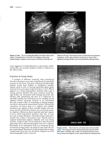
around the foreign body can easily be seen until the air/ Figure 3.121. This horse had a draining tract at the shoulder
gas is introduced. If retrieval of the foreign body is more region. There appears to be a dense foreign body several centim
complicated, then the area should be mapped out with eters deep, consistent with a bullet. The surgical approach to the
ultrasonography before retrieval. foreign body was mapped out with ultrasonography intraoperatively.