
Page 373 - Adams and Stashak's Lameness in Horses, 7th Edition
P. 373
Diagnostic Imaging 339
VetBooks.ir
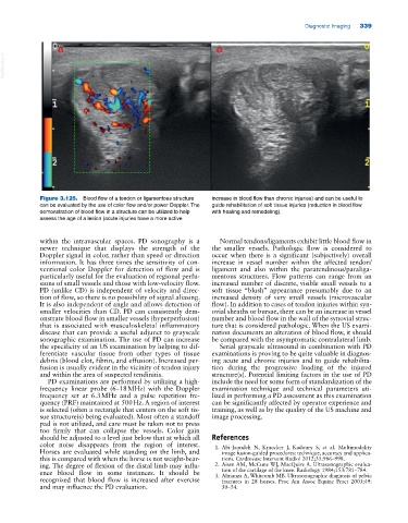
Figure 3.125. Blood flow of a tendon or ligamentous structure increase in blood flow than chronic injuries) and can be useful to
can be evaluated by the use of color flow and/or power Doppler. The guide rehabilitation of soft tissue injuries (reduction in blood flow
demonstration of blood flow in a structure can be utilized to help with healing and remodeling).
assess the age of a lesion (acute injuries have a more active
within the intravascular spaces. PD sonography is a Normal tendons/ligaments exhibit little blood flow in
newer technique that displays the strength of the the smaller vessels. Pathologic flow is considered to
Doppler signal in color, rather than speed or direction occur when there is a significant (subjectively) overall
information. It has three times the sensitivity of con increase in vessel number within the affected tendon/
ventional color Doppler for detection of flow and is ligament and also within the paratendinous/paraliga
particularly useful for the evaluation of regional perfu mentous structures. Flow patterns can range from an
sions of small vessels and those with low‐velocity flow. increased number of discrete, visible small vessels to a
PD (unlike CD) is independent of velocity and direc soft tissue “blush” appearance presumably due to an
tion of flow, so there is no possibility of signal aliasing. increased density of very small vessels (microvascular
It is also independent of angle and allows detection of flow). In addition to cases of tendon injuries within syn
smaller velocities than CD. PD can consistently dem ovial sheaths or bursae, there can be an increase in vessel
onstrate blood flow in smaller vessels (hyperperfusion) number and blood flow in the wall of the synovial struc
that is associated with musculoskeletal inflammatory ture that is considered pathologic. When the US exami
disease that can provide a useful adjunct to grayscale nation documents an alteration of blood flow, it should
sonographic examination. The use of PD can increase be compared with the asymptomatic contralateral limb.
the specificity of an US examination by helping to dif Serial grayscale ultrasound in combination with PD
ferentiate vascular tissue from other types of tissue examinations is proving to be quite valuable in diagnos
debris (blood clot, fibrin, and effusion). Increased per ing acute and chronic injuries and to guide rehabilita
fusion is usually evident in the vicinity of tendon injury tion during the progressive loading of the injured
and within the area of suspected tendinitis. structure(s). Potential limiting factors in the use of PD
PD examinations are performed by utilizing a high‐ include the need for some form of standardization of the
frequency linear probe (6–18 MHz) with the Doppler examination technique and technical parameters uti
frequency set at 6.3 MHz and a pulse repetition fre lized in performing a PD assessment as this examination
quency (PRF) maintained at 500 Hz. A region of interest can be significantly affected by operator experience and
is selected (often a rectangle that centers on the soft tis training, as well as by the quality of the US machine and
sue structure(s) being evaluated). Most often a standoff image processing.
pad is not utilized, and care must be taken not to press
too firmly that can collapse the vessels. Color gain
should be adjusted to a level just below that at which all References
color noise disappears from the region of interest. 1. Abi‐Jaoudeh N, Kruecker J, Kadoury S, et al. Multimodality
Horses are evaluated while standing on the limb, and image fusion‐guided procedures: technique, accuracy and applica
this is compared with when the horse is not weight‐bear tions. Cardiovasc Intervent Radiol 2012;35:986–998.
ing. The degree of flexion of the distal limb may influ 2. Aisen AM, McCune WJ, MacQuire A. Ultrasonographic evalua
ence blood flow in some instances. It should be tion of the cartilage of the knee. Radiology 1984;153:781–784.
recognized that blood flow is increased after exercise 3. Almanza A, Whitcomb MB. Ultrasonographic diagnosis of pelvic
fractures in 28 horses. Proc Am Assoc Equine Pract 2003;49:
and may influence the PD evaluation. 50–54.