
Page 378 - Adams and Stashak's Lameness in Horses, 7th Edition
P. 378
344 Chapter 3
Table 3.1. Suggested minimum image acquisition counts.
VetBooks.ir Body region Number of counts (×1,000)
Foot
100–150
Carpus 100–150
Elbow 150–200
Shoulder 200–300
Tarsus 150–200
Stifle 150–200
Sacroiliac area 200–300
Spine 200–300
Soft tissue (pool) phase image 75–100
Motion correction acquisition 2 seconds/frame for 40–50
frames
Image acquisition is determined either by the number
of counts or the acquisition time. The greater the number
of counts/image, the better the image quality, excluding
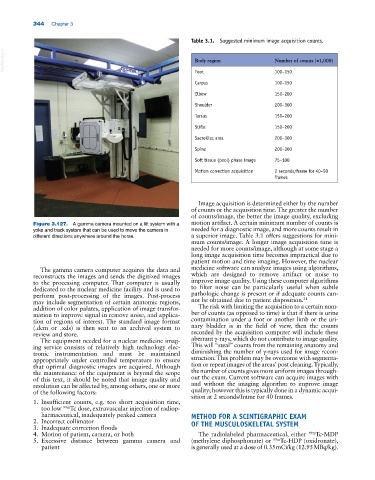
Figure 3.127. A gamma camera mounted on a lift system with a motion artifact. A certain minimum number of counts is
yoke and track system that can be used to move the camera in needed for a diagnostic image, and more counts result in
different directions anywhere around the horse. a superior image. Table 3.1 offers suggestions for mini
mum counts/image. A longer image acquisition time is
needed for more counts/image, although at some stage a
long image acquisition time becomes impractical due to
patient motion and time imaging. However, the nuclear
The gamma camera computer acquires the data and medicine software can analyze images using algorithms,
reconstructs the images and sends the digitized images which are designed to remove artifact or noise to
to the processing computer. That computer is usually improve image quality. Using these computer algorithms
dedicated to the nuclear medicine facility and is used to to filter noise can be particularly useful when subtle
perform post‐processing of the images. Post‐process pathologic change is present or if adequate counts can
may include segmentation of certain anatomic regions, not be obtained due to patient disposition. 31
addition of color palates, application of image transfor The risk with limiting the acquisition to a certain num
mation to improve signal in remove noise, and applica ber of counts (as opposed to time) is that if there is urine
tion of regions of interest. The standard image format contamination under a foot or another limb or the uri
(.dcm or .xds) is then sent to an archival system to nary bladder is in the field of view, then the counts
review and store. recorded by the acquisition computer will include these
The equipment needed for a nuclear medicine imag aberrant γ‐rays, which do not contribute to image quality.
ing service consists of relatively high technology elec This will “steal” counts from the remaining anatomy and
tronic instrumentation and must be maintained diminishing the number of γ‐rays used for image recon
appropriately under controlled temperature to ensure struction. This problem may be overcome with segmenta
that optimal diagnostic images are acquired. Although tion or repeat images of the areas’ post cleaning. Typically,
the maintenance of the equipment is beyond the scope the number of counts gives more uniform images through
of this text, it should be noted that image quality and out the exam. Current software can acquire images with
resolution can be affected by, among others, one or more and without the imaging algorithm to improve image
of the following factors: quality; however this is typically done in a dynamic acqui
sition at 2 seconds/frame for 40 frames.
1. Insufficient counts, e.g. too short acquisition time,
too low 99m Tc dose, extravascular injection of radiop
harmaceutical, inadequately peaked camera METHOD FOR A SCINTIGRAPHIC EXAM
2. Incorrect collimator OF THE MUSCULOSKELETAL SYSTEM
3. Inadequate correction floods
4. Motion of patient, camera, or both The radiolabeled pharmaceutical, either 99m Tc‐MDP
5. Excessive distance between gamma camera and (methylene diphosphonate) or 99m Tc‐HDP (oxidronate),
patient is generally used at a dose of 0.35 mCi/kg (12.95 MBq/kg).