
Page 383 - Adams and Stashak's Lameness in Horses, 7th Edition
P. 383
Diagnostic Imaging 349
VetBooks.ir
Figure 3.134. Composite image of blood flow in the region of
the caudal aorta, showing normal bifurcation of the aorta (arrow) Figure 3.135. Soft tissue (pool) phase view of a normal left
into internal and external iliac arteries. forefoot, showing vascular activity on the palmar aspects proximal
and distal to the fetlock and in the area of the coronary band
(arrows). Note that the fetlock and pastern regions have
homogeneously smooth uptake.
A B
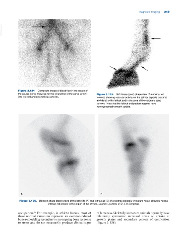
Figure 3.136. Delayed phase lateral views of the left stifle (A) and left tarsus (B) of a normal skeletally immature horse, showing normal
intense radiotracer in the region of the physes. Source: Courtesy of Dr. Erik Bergman.
occupation. For example, in athletic horses, most of of lameness. Skeletally immature animals normally have
96
these normal variations represent an exercise‐induced bilaterally symmetric increased areas of uptake at
bone remodeling secondary to an ongoing bone response growth plates and secondary centers of ossification
to stress and do not necessarily produce clinical signs (Figure 3.136).