
Page 386 - Adams and Stashak's Lameness in Horses, 7th Edition
P. 386
VetBooks.ir
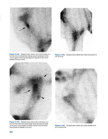
Figure 3.143. Delayed phase lateral view of the shoulder of a Figure 3.145. Delayed phase lateral view of the elbow joint of a
normal horse. A normal focal intense area of radiotracer on the normal horse.
cranial aspect of the proximal diaphysis represents the normal
deltoid tuberosity (arrow).
Figure 3.144. Delayed phase lateral view of the femur of a
normal horse. Note the cranial (large arrow) and caudal (small
arrow) parts of the greater trochanter and the third trochanter Figure 3.146. Delayed phase lateral view of the shoulder joint
(arrowhead) as separate structures. of a normal horse.
352