
Page 387 - Adams and Stashak's Lameness in Horses, 7th Edition
P. 387
Diagnostic Imaging 353
VetBooks.ir
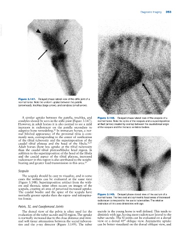
Figure 3.147. Delayed phase lateral view of the stifle joint of a
normal horse. Note the uniform uptake between the patella
(arrowhead), trochlea (large arrow), and condyles (small arrow).
A similar uptake between the patella, trochlea, and Figure 3.148. Delayed phase lateral view of the scapula of a
condyles should be seen in the stifle joint (Figure 3.147). normal horse. Note the spine of the scapula and a superimposition
However, in adult horses it is also normal to see a mild artifact (arrow) created by overlap between the caudodorsal angle
increases in radiotracer on the patella secondary to of the scapula and the thoracic vertebral bodies.
26
adaptive bone remodeling. In immature horses, a nor
mal bilobed appearance of the proximal tibia is com
monly seen, corresponding to the center of ossification
of the tibial tuberosity and the superimposition of the
caudal tibial plateau and the head of the fibula. 26,71
Adult horses show less uptake at the tibial tuberosity
than the caudal tibial plateau/fibular head region. In
addition to the superimposition of the head of the fibula
and the caudal aspect of the tibial plateau, increased
radiotracer in this region is also attributed to the weight‐
bearing and greater load transmission in this area. 26
Scapula
The scapula should be easy to visualize, and in some
cases the withers can be evaluated at the same time
(Figure 3.148). Superimposition artifact from the with
ers and thoracic spine often occurs on images of the
scapula, creating an area of perceived increased uptake.
The caudal border and the spine of the scapula have
normally greater uptake than the supra‐ and infraspina Figure 3.149. Delayed phase dorsal view of the sacrum of a
tus fossae. normal horse. The two oval and symmetric focal areas of increased
radiotracer correspond to the sacral tuberosities. The relative
distinction of this area diminishes with age.
Pelvis, SI, and Coxofemoral Joints
The dorsal view of the pelvis is often used for the sacrale in the young horse is well defined. This tends to
evaluation of the tuber sacrale and SI region. The uptake diminish with age, having more radiotracer lateral to the
is normally increased due to the close distance and mini tuber sacrale. The SI joints can be evaluated on a dorsal
mal soft tissue attenuation between the sacral tuberosi view or a dorsal 45° oblique view. Asymmetric uptake
ties and the γ‐ray detector (Figure 3.149). The tuber can be better visualized on the dorsal oblique view, and