
Page 385 - Adams and Stashak's Lameness in Horses, 7th Edition
P. 385
Diagnostic Imaging 351
VetBooks.ir
A B
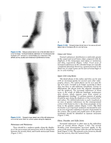
Figure 3.142. Delayed phase dorsal view of the carpus (A) and
lateral view of the tarsus (B) of a normal horse.
Figure 3.140. Delayed phase lateral view of the left distal limb of
a normal horse. The focal moderate radiotracer on the dorsoproximal Carpus and Tarsus
diaphysis of P1 (arrow) is a normal finding as a response to the
athletic activity usually seen bilaterally in performance horses. Normal radiotracer distribution is uniformly greater
in the carpal and tarsal bones when compared with the
diaphysis of the metacarpus and radius or metatarsus
and tibia, respectively (Figure 3.142). Focal areas of
increased radiotracer in the cuboidal bones are typically
considered abnormal. However, a general increase in the
distal row of carpal bones can be seen as horses begin or
are in early training. 35
Upper Limb Long Bones
The distal physis of the radius and tibia can be seen
with an increased uptake for several years after radio
graphic closure. The radial physis can be seen past the
age of 8 years. Growth is no longer occurring at these
sites, but there is still sufficient osteoblastic activity to
differentiate the physis from the adjacent metaphysis
and the epiphysis. The increased radiotracer in those
areas is the result of different histologic architecture
after closure, which exposes more bone crystal to
diphosphonate binding. A uniform pattern of uptake
96
should be seen along the diaphysis of the normal tibia
97
and radius. The deltoid tuberosity is easily visualized as
an area of greater radiotracer on the cranioproximal
cortex of the humerus due to the closer proximity of the
bone to the gamma camera (Figure 3.143). The third
trochanter is an important landmark and should be seen
as an area of greater radiotracer because of proximity to
the camera. The cranial and caudal parts of the greater
trochanter should be identified as separate structures
(Figure 3.144).
Figure 3.141. Delayed phase lateral view of the left metacarpus
of a normal horse. Note the uniform uptake along the diaphysis.
Elbow, Shoulder, and Stifle Joints
Increased radiotracer is often seen in the radioulnar
Metacarpus and Metatarsus joint of normal elbows (Figure 3.145). The normal
There should be a uniform uptake along the diaphy shoulder joint demonstrates increased uptake in the
sis of the metacarpus and metatarsus with no distinction areas of the greater and lesser tubercles and the humeral
between the second, third, and fourth metacarpal bones head (Figure 3.146). The glenoid cavity, however, should
(Figure 3.141). have less activity than the humeral head.