
Page 380 - Adams and Stashak's Lameness in Horses, 7th Edition
P. 380
346 Chapter 3
The following views are recommended for the full dorsal pelvis, caudal pelvis (tail on detector, TOD), and
evaluation of the thoracic and pelvic limbs and spine RDO/LDO sacrum. Lateral and dorsal views of the
VetBooks.ir Thoracic Limb the camera is from the vertebra. They can, however, be
spine are less effective due to the great distance that
using a detector with a 20‐inch (50‐cm) field of view.
included as orthogonal views when a lesion is found.
Lateral and dorsal views of the foot (these views A point source, also known as fiducial marker, (e.g.
cobalt marker or the syringe and needle that was used
include distal metacarpus, metacarpophalangeal joint, for the 99m Tc‐HDP injection, sealed in a latex glove) can
and phalanges), solar distal phalanx, lateral and dorsal be placed along the dorsum of the back to localize the
carpus, lateral and cranial humerus, and lateral scapula. exact position of a lesion, which is then marked with a
The metacarpus is included in the views of the foot and permanent marker.
the carpus, the radius and elbows are included in the
views of the carpus and humerus, and the shoulder is
included in the views of the humerus and scapula. If an Foot
area of abnormal uptake is seen in the metacarpus, radius, Different options exist for obtaining lateral, dorsal,
or elbow, or in an area where only lateral views were and palmar views of the feet. In some institutions, the
obtained, additional images including orthogonal views nuclear medicine suite has a pit in the floor into which
centered over the areas of interest should be acquired. the gamma camera is lowered to have it centered over
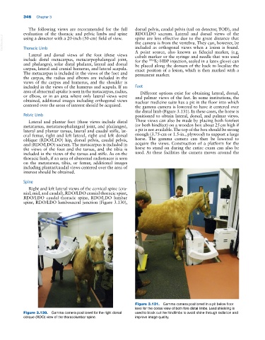
the distal limb (Figure 3.131). In these cases, the horse is
Pelvic Limb positioned to obtain lateral, dorsal, and palmar views.
Lateral and plantar foot (these views include distal These views can also be made by placing both forefeet
metatarsus, metatarsophalangeal joint, and phalanges), (or both hindfeet) on a wooden box about 25 cm high if
lateral and plantar tarsus, lateral and caudal stifle, lat a pit is not available. The top of the box should be strong
eral femur, right and left lateral, right and left dorsal enough (3.75‐cm or 1.5‐in., plywood) to support a large
oblique (RDO/LDO) hip, dorsal pelvis, caudal pelvis, horse. The gamma camera can then be lowered to
and (RDO/LDO) sacrum. The metacarpus is included in acquire the views. Construction of a platform for the
the views of the foot and the tarsus, and the tibia is horse to stand on during the entire exam can also be
included in the views of the tarsus and stifle. As on the used. At these facilities the camera moves around the
thoracic limb, if an area of abnormal radiotracer is seen
on the metatarsus, tibia, or femur, additional images
including plantar/caudal views centered over the area of
interest should be obtained.
Spine
Right and left lateral views of the cervical spine (cra
nial, mid, and caudal), RDO/LDO cranial thoracic spine,
RDO/LDO caudal thoracic spine, RDO/LDO lumbar
spine, RDO/LDO lumbosacral junction (Figure 3.130),
Figure 3.131. Gamma camera positioned in a pit below floor
level for the dorsal view of both fore distal limbs. Lead shielding is
Figure 3.130. Gamma camera positioned for the right dorsal used to block out the hindlimbs to avoid shine through radiation and
oblique (RDO) view of the thoracolumbar spine. improve image quality.