
Page 381 - Adams and Stashak's Lameness in Horses, 7th Edition
P. 381
Diagnostic Imaging 347
VetBooks.ir
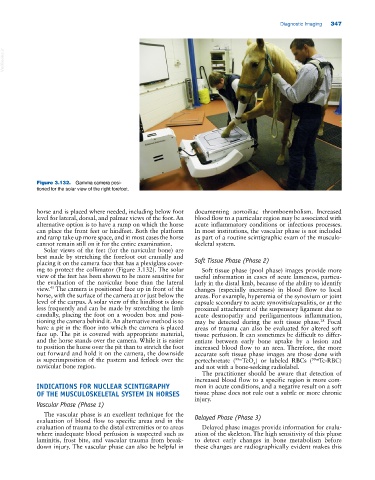
Figure 3.132. Gamma camera posi
tioned for the solar view of the right forefoot.
horse and is placed where needed, including below foot documenting aortoiliac thromboembolism. Increased
level for lateral, dorsal, and palmar views of the foot. An blood flow to a particular region may be associated with
alternative option is to have a ramp on which the horse acute inflammatory conditions or infectious processes.
can place the front feet or hindfeet. Both the platform In most institutions, the vascular phase is not included
and ramp take up more space, and in most cases the horse as part of a routine scintigraphic exam of the musculo
cannot remain still on it for the entire examination. skeletal system.
Solar views of the feet (for the navicular bone) are
best made by stretching the forefoot out cranially and
placing it on the camera face that has a plexiglass cover Soft Tissue Phase (Phase 2)
ing to protect the collimator (Figure 3.132). The solar Soft tissue phase (pool phase) images provide more
view of the feet has been shown to be more sensitive for useful information in cases of acute lameness, particu
the evaluation of the navicular bone than the lateral larly in the distal limb, because of the ability to identify
45
view. The camera is positioned face up in front of the changes (especially increases) in blood flow to local
horse, with the surface of the camera at or just below the areas. For example, hyperemia of the synovium or joint
level of the carpus. A solar view of the hindfoot is done capsule secondary to acute synovitis/capsulitis, or at the
less frequently and can be made by stretching the limb proximal attachment of the suspensory ligament due to
caudally, placing the foot on a wooden box and posi acute desmopathy and periligamentous inflammation,
tioning the camera behind it. An alternative method is to may be detected during the soft tissue phase. Focal
28
have a pit in the floor into which the camera is placed areas of trauma can also be evaluated for altered soft
face up. The pit is covered with appropriate material, tissue perfusion. It can sometimes be difficult to differ
and the horse stands over the camera. While it is easier entiate between early bone uptake by a lesion and
to position the horse over the pit than to stretch the foot increased blood flow to an area. Therefore, the more
out forward and hold it on the camera, the downside accurate soft tissue phase images are those done with
is superimposition of the pastern and fetlock over the pertechnetate ( 99m TcO ) or labeled RBCs ( 99m Tc‐RBC)
4
navicular bone region. and not with a bone‐seeking radiolabel.
The practitioner should be aware that detection of
increased blood flow to a specific region is more com
INDICATIONS FOR NUCLEAR SCINTIGRAPHY mon in acute conditions, and a negative result on a soft
OF THE MUSCULOSKELETAL SYSTEM IN HORSES tissue phase does not rule out a subtle or more chronic
injury.
Vascular Phase (Phase 1)
The vascular phase is an excellent technique for the
evaluation of blood flow to specific areas and in the Delayed Phase (Phase 3)
evaluation of trauma to the distal extremities or to areas Delayed phase images provide information for evalu
where inadequate blood perfusion is suspected such as ation of the skeleton. The high sensitivity of this phase
laminitis, frost bite, and vascular trauma from break to detect early changes in bone metabolism before
down injury. The vascular phase can also be helpful in these changes are radiographically evident makes this