
Page 371 - Adams and Stashak's Lameness in Horses, 7th Edition
P. 371
Diagnostic Imaging 337
Elastography to Evaluate Soft Tissue Injuries of acute injuries with elastography can reveal signifi
cantly increased softness in areas of hematoma and
Elastography is a new ultrasonographic technique
VetBooks.ir that allows an examiner to evaluate the mechanical detect the tissue becoming progressively stiffer with
fiber disruption. Serial elastographic examinations can
properties of soft tissues.
This technique estimates
8,51,67
healing.
tissue strain, where strain is defined as the fractional
An initial study was performed to determine the fea
change in length of a tissue when an external force is sibility, reproducibility, and repeatability of elastogra
applied. Elastography evaluates tissue motion as phy and to establish the normal elastographic appearance
compression is applied to the tissue using manual of the tendons of the equine metacarpus. A group of
52
compression with the ultrasound transducer. The tissue sound horses without evidence of musculoskeletal
displacement causes a measureable displacement of the pathology were evaluated with standard grayscale ultra
ultrasound waves. Soft, deformable structures, such as sound, and elastographic evaluations were performed
damaged or inflamed tissues, cause greater displacement using an ultrasound scanner (MyLab 70 ultrasound
of sound waves than hard, rigid structures. Elastography system Biosound Esaote Inc. Indianapolis, IN) with a
can therefore provide information about the behavior of 6–18‐MHz linear probe using a previously described
internal soft tissue structures when compressed. Since technique. Elastographic images of the SDF and DDF
pathologic tissues often exhibit altered mechanical tendons and the branches of the SL were evaluated
properties, elastography allows the differentiation quantitatively and qualitatively.
between normal and diseased tissues due to the differ An extension of that study in normal horse was per
ences in these elastic properties. With elastography, formed in horses with clinical conditions found with
compression (in a transverse or longitudinal plane) is grayscale ultrasound. This study attempted to character
applied with the ultrasound probe, and tissue displace ize elastographic findings of clinical cases with soft tis
ment is measured by comparing the echo signals before sue injuries of the distal limb and to evaluate the
and after compression. This is in contrast to conven differences in the elastographic appearance of acute ver
tional grayscale ultrasonography that only evaluates tis sus chronic injuries. Horses were evaluated through
53
sue morphology and the differences between acoustic out their rehabilitation and assessed for changes in the
impedance of adjacent structures. ultrasonographic and elastographic appearance over
Elastography was initially used in humans to help dif time. These findings were correlated with clinical heal
ferentiate benign from malignant nodules in the breast, ing. This study found that compression elastography
prostate, thyroid, liver, and lymph tissue and for evalu corresponded well with clinical evaluation using gray
ating hepatic fibrosis. Recently, elastography was used scale ultrasound (Figure 3.124). Acute lesions were
67
to evaluate this technology in musculoskeletal injuries in significantly softer than subacute/chronic lesions when
humans primarily the Achilles tendon. 29,30 The evaluation
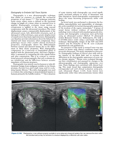
Figure 3.124. Elastography, a new software program available in some advanced ultrasound systems that can measure the strain within
that structure and portray the softness of a structure displayed by a difference with color codes.