
Page 366 - Adams and Stashak's Lameness in Horses, 7th Edition
P. 366
332 Chapter 3
resolution over time, which can aid surgical decision‐
making, direct management, and rehabilitation strategies
VetBooks.ir er’s ability to accurately diagnose and manage joint‐
and increase the understanding of joint disease in horses.
Ultrasonography can enhance the equine practition
related problems in performance horses. However, it is
only one tool at the clinician’s disposal; clinical acumen
and selective use of other imaging modalities are needed
to accurately diagnose many types of joint disease.
Currently, the gold standard for assessing soft tissue
injuries in humans is MRI. However, in human medi
cine, ultrasonography remains a very useful imaging
modality for evaluating the popliteal space, knee, patel
lar tendon, shoulder (especially the rotator cuff), and
neonatal hip. Extensive use of ultrasonography in the
examination of equine joints has already demonstrated
the sensitivity of this modality in defining articular
lesions not apparent radiographically. In time, there will
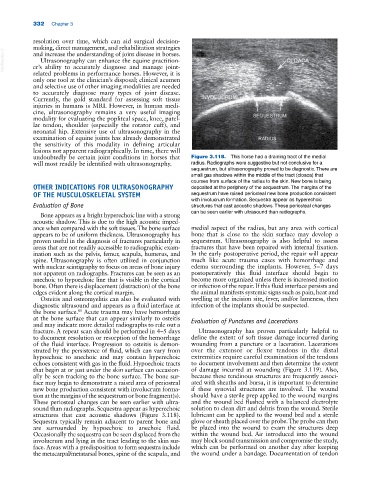
undoubtedly be certain joint conditions in horses that Figure 3.118. This horse had a draining tract of the medial
will most readily be identified with ultrasonography. radius. Radiographs were suggestive but not conclusive for a
sequestrum, but ultrasonography proved to be diagnostic. There are
small gas shadows within the middle of the tract (cloaca) that
courses from surface of the radius to the skin. New bone is being
OTHER INDICATIONS FOR ULTRASONOGRAPHY deposited at the periphery of the sequestrum. The margins of the
OF THE MUSCULOSKELETAL SYSTEM sequestrum have raised periosteal new bone production consistent
with involucrum formation. Sequestra appear as hyperechoic
Evaluation of Bone structures that cast acoustic shadows. These periosteal changes
can be seen earlier with ultrasound than radiographs.
Bone appears as a bright hyperechoic line with a strong
acoustic shadow. This is due to the high acoustic imped
ance when compared with the soft tissues. The bone surface medial aspect of the radius, but any area with cortical
appears to be of uniform thickness. Ultrasonography has bone that is close to the skin surface may develop a
proven useful in the diagnosis of fractures particularly in sequestrum. Ultrasonography is also helpful to assess
areas that are not readily accessible to radiographic exam fractures that have been repaired with internal fixation.
ination such as the pelvis, femur, scapula, humerus, and In the early postoperative period, the repair will appear
spine. Ultrasonography is often utilized in conjunction much like acute trauma cases with hemorrhage and
with nuclear scintigraphy to focus on areas of bone injury edema surrounding the implants. However, 5–7 days
not apparent on radiographs. Fractures can be seen as an postoperatively this fluid interface should begin to
anechoic to hypoechoic line that is visible in the cortical become more organized unless there is increased motion
bone. Often there is displacement (distraction) of the bone or infection of the repair. If this fluid interface persists and
edges evident along the cortical margin. the animal manifests systemic signs such as pain, heat and
Osteitis and osteomyelitis can also be evaluated with swelling at the incision site, fever, and/or lameness, then
diagnostic ultrasound and appears as a fluid interface at infection of the implants should be suspected.
the bone surface. Acute trauma may have hemorrhage
81
at the bone surface that can appear similarly to osteitis Evaluation of Punctures and Lacerations
and may indicate more detailed radiographs to rule out a
fracture. A repeat scan should be performed in 4–5 days Ultrasonography has proven particularly helpful to
to document resolution or resorption of the hemorrhage define the extent of soft tissue damage incurred during
of the fluid interface. Progression to osteitis is demon wounding from a puncture or a laceration. Lacerations
strated by the persistence of fluid, which can vary from over the extensor or flexor tendons in the distal
hypoechoic to anechoic and may contain hyperechoic extremities require careful examination of the tendons
echoes consistent with gas in the fluid. Hypoechoic tracts to document involvement and then determine the extent
that begin at or just under the skin surface can occasion of damage incurred at wounding (Figure 3.119). Also,
ally be seen tracking to the bone surface. The bone sur because these tendinous structures are frequently associ
face may begin to demonstrate a raised area of periosteal ated with sheaths and bursa, it is important to determine
new bone production consistent with involucrum forma if these synovial structures are involved. The wound
tion at the margins of the sequestrum or bone fragment(s). should have a sterile prep applied to the wound margins
These periosteal changes can be seen earlier with ultra and the wound bed flushed with a balanced electrolyte
sound than radiographs. Sequestra appear as hyperechoic solution to clean dirt and debris from the wound. Sterile
structures that cast acoustic shadows (Figure 3.118). lubricant can be applied to the wound bed and a sterile
Sequestra typically remain adjacent to parent bone and glove or sheath placed over the probe. The probe can then
are surrounded by hypoechoic to anechoic fluid. be placed into the wound to exam the structures deep
Occasionally the sequestra can be seen displaced from the within the wound bed. Air introduced into the wound
involucrum and lying in the tract leading to the skin sur may block sound transmission and compromise the study,
face. Areas with a predisposition to form sequestra include which can be performed on another day after keeping
the metacarpal/metatarsal bones, spine of the scapula, and the wound under a bandage. Documentation of tendon