
Page 357 - Adams and Stashak's Lameness in Horses, 7th Edition
P. 357
Diagnostic Imaging 323
and ultrasound will identify specific indications for each
imaging modality and improve our capabilities as
VetBooks.ir
ultrasonographers.
ULTRASOUND TO EVALUATE JOINT INJURY
Joint injury, osteochondrosis, and degenerative joint
disease are significant causes of lameness in the horse. 68,69
An accurate diagnosis of the cause of joint pain can
prove critical for selecting the most appropriate
treatment(s) and rehabilitation. Intra‐articular diagnos
tic anesthesia should be utilized to localize a lameness to
a particular joint. Radiology, ultrasonography, thermog
raphy, nuclear imaging, CT, and MRI all have a place in
lameness diagnostics, and each may be warranted in
selected cases. 50,62 However, many of the joint problems
seen in the horse can be effectively imaged with a com
bination of radiographs and ultrasound. Good‐quality
baseline images are critical to provide a diagnosis but
also will be helpful in reevaluation of the joint in the
future. The radiographic exam is most effective at evalu
ating the bony structure of the joints of the equine limb.
In some joints, there can be poor correlation between
clinical and radiographic findings, or the radiographic
study may be inconclusive. 55,62 In these types of cases the
cause of lameness is presumed to be soft tissue injury,
and often empirical treatment is instituted. A complete
ultrasonographic exam is indicated in these cases to
evaluate the periarticular tendons and ligaments, the
joint capsule, and the joint fluid, as well as to provide
valuable information about the cartilage and subchon
dral bone surface. 2,17–20,58,78,79 Ultrasonography has the
added advantage of providing immediate, detailed infor
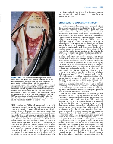
Figure 3.111. The structures within the digital flexor tendon mation through a range of motion (in extension and
sheath (DFTS) are exposed via a longitudinal incision through the flexion). However, ultrasound provides a relatively lim
annular ligament and the SDFT at the level of the fetlock joint. The ited field of view when compared with other imaging
unique anatomy at this level begins with the mesotendinous techniques making global survey of a joint to be time
attachments (not seen in this section) to the DDFT at the medial and consuming. When used in combination, radiography
lateral borders in the proximal sheath. Progressing distally the SDFT and ultrasonography are complementary and can sup
produces the proximal ring of the manica flexoria encircling the DDFT. ply more information about an orthopedic problem
Just distal to the manica flexoria, the SDFT and DDFT are bound than when each modality is used alone.
within the fetlock canal by the primary annular ligament (PAL), which The clinician must understand the advantages and
has been incised in this section. Just distal to the fetlock canal, the
SDFT makes another ring (smaller) around the DDFT called the limitations of each imaging modality as well as the
distal ring of the manica flexoria. Just distal to this ring, the vincula appearance of the normal anatomy and pathophysiology
attach to the midline of the DDFT on its dorsal border. characteristic with each technique. When utilizing
diagnostic ultrasound to evaluate joints, it is necessary
to become familiar with the appearance of many of
MRI examination. While ultrasonography and MRI these structures in transverse, longitudinal (sagittal), and
remain the optimal choices for soft tissue imaging, it frontal planes. Normal anatomical variations exist, and
remains unclear in all cases which is the most effective it is important that the clinician recognizes these varia
imaging tool. In humans there are many studies that tions as normal to prevent developing an inaccurate
have been performed that compare the diagnostic accu diagnosis. The opposite limb can and should be used for
racy and utility of ultrasound compared with MRI for a comparison when possible recognizing that disease can
variety of orthopedic problems. These studies are lack occur at the same location in the opposite limb.
ing in horses and need to be performed. A limited num In general, the stability of a joint is provided by the
ber of studies have been performed and have looked at congruent contours of the joint surfaces of the bony
the collateral ligaments of the distal interphalangeal column, the collateral (and in some joints the intra‐articular)
joint and the proximal plantar region. 6,35,36 In those ligaments, the integrity of the joint capsule, and the
studies a negative ultrasound study does not rule out an surrounding tendons and ligaments. In the proximal
abnormality and positive ultrasonographic needs to be limb, various muscle groups and tendons that cross the
regarded with caution. It is hoped that further experi joints provide additional stability. Most joints of the
ence comparing ultrasound with MRI along with the appendicular skeleton in the horse are designed to work in
completion of more comparative studies between MRI the sagittal plane with flexion and extension being the