
Page 350 - Adams and Stashak's Lameness in Horses, 7th Edition
P. 350
316 Chapter 3
VetBooks.ir
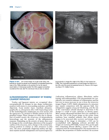
B
A
C
Figure 3.101. (A) To best image the origin of the OSLs, the longitudinally to image the origin of the OSLs to their respective
transducer should be placed more abaxially and directed toward the PSBs. The longitudinal examination should image the insertion of
base of the PSBs (similarly to the position for the SL branch the OSL onto the proximal sesamoid bone (C). Source: US images
examination). In transverse section the OSLs appear somewhat courtesy of Dr. Caitlyn Horne.
rounded at this level. (B) The transducer should also be placed
ULTRASONOGRAPHIC ASSESSMENT OF TENDON/ (indicating inflammation, edema, fibroplasia, and/or
LIGAMENT PATHOLOGY tearing) and enthesopathies. CSA measurements are con
sidered a very sensitive indicator of inflammation, and the
Tendon and ligament injuries are recognized ultra best way to assess increases in size is from the transverse
sonographically by changes in size, shape, architecture, images (Figure 3.109). Subtle enlargements of a structure
position (with respect to surrounding anatomy), and fiber may require comparison with the opposite limb. Any
alignment. Acute tendon injury manifests ultrasono enlargement suggests structural thickening, and the rest
graphically as enlargement often with hypoechogenicity of the examination should attempt to determine if the
of the tendon/ligament architecture on cross section and change is a result of acute, subacute, or chronic injury.
the appearance of a striated or fibrillar pattern in the lon Most current ultrasound machines have the capability to
gitudinal images. These changes are often due to disrup trace the CSA of the frozen image on the screen. Some
tion of tendon matrix, the occurrence of intratendinous machines have installed software that allows stored
hemorrhage, and the intensity of the inflammatory images to be processed after recovery and storage. Post‐
response. Depending on the severity of injury, there may processing of the image to determine a CSA can be
be changes in the overall shape, distortion of the margins/ accomplished with digitizing software provided the
outline of the structure, and a change in the normal ana images are stored as DICOMs; however this can be quiet
tomical position in relation to other structures in that time consuming (Dicom Works ). Lesion CSA can be
R
area. The most consistently diagnosed tendon and liga compared with the tendon/ligament CSA measurement to
ment injuries include enlargement and hypoechogenicity calculate the proportion of tendon involved. An accurate