
Page 469 - Adams and Stashak's Lameness in Horses, 7th Edition
P. 469
Diagnostic Imaging 435
99.1 °F
A
B
VetBooks.ir 95
90
85
80
79.1
99.0 °F 99.5 °F
C D
95 95
90
90
85 85
80 80
79.0 79.5
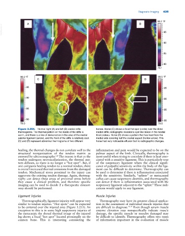
Figure 3.254. Normal right (A) and left (B) cranial stifle horses. Horse (C) shows a focal hot spot (circle) over the distal
thermograms. The thermal pattern on the inside of the stifle is medial stifle; radiographs revealed a cyst‐like lesion in the medial
warm, and there is a line of demarcation in the area of the medial tibial plateau. Horse (D) shows a pattern that has heat from the
patellar ligament (arrow), and the front of the stifle is relatively cool. medial side covering half the cranial aspect (hollow arrow). This
(C) and (D) represent abnormal thermograms of two different horse had very noticeable effusion but no radiographic changes.
healing, the thermal changes do not correlate well to the inflammation and pain would be expected to be on the
structural reorganization of the tendon matrix as palmar aspect of the limb. Clinically, thermography is
assessed by ultrasonography. The reason is that as the most useful when trying to correlate if there is heat asso
2,4
tendon undergoes neovascularization, the thermal pat ciated with a sensitive ligament. This is particularly true
tern diffuses, so there is no longer a “hot spot”. But, if of the suspensory ligament where the clinical signifi
one compares healing tendon to a normal tendon, there cance of palpable sensitivity within the body of the liga
is overall increased thermal emissions from the damaged ment can be difficult to determine. Thermography can
tendon. Mechanical stress proximal to the injury can be used to determine if there is inflammation associated
aggravate the existing tendon damage. Again, thermog with the sensitivity. Similarly, “splints” or metacarpal
raphy can detect these areas of proximal stress before callus can cause suspensory desmitis, and thermography
they cause a clinical problem, and therefore specific can detect if there is inflammation associated with the
imaging can be used to decide if a therapeutic desmot suspensory ligament adjacent to the “splint.” These indi
omy should be performed. cations would apply to any ligament.
Ligament Injuries Muscle Injuries
Thermographically, ligament injuries will appear very Thermography may have its greatest clinical applica
similar to tendon injuries. “Hot spots” can be expected tion in the assessment of individual muscle injuries that
to be centered over the injured area (Figure 3.255). An are difficult to diagnose. 14,19 Even though serum muscle
exception to this is in some high suspensory injuries of enzyme elevation may nonspecifically indicate muscle
the metacarpi; the dorsal thermal image of the injured damage, the specific muscle or muscles damaged may
leg shows a focal “hot spot” located proximally on the be difficult to identify. Thermography offers two types
cannon bone. This is interesting considering the of information important in the evaluation of muscle