
Page 470 - Adams and Stashak's Lameness in Horses, 7th Edition
P. 470
436 Chapter 3
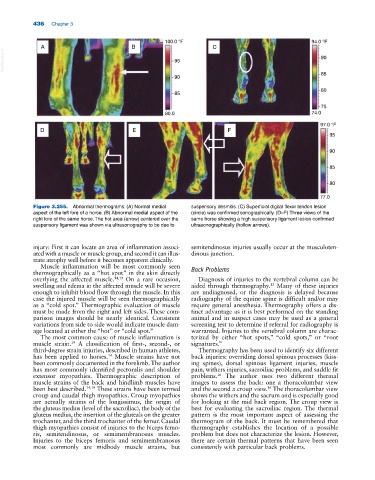
100.0 °F 94.0 °F
A B C
VetBooks.ir 95 90
90 85
80
85
75
80.0 74.0
97.0 °F
D E F
95
90
85
80
77.0
Figure 3.255. Abnormal thermograms. (A) Normal medial suspensory desmitis. (C) Superficial digital flexor tendon lesion
aspect of the left fore of a horse. (B) Abnormal medial aspect of the (circle) was confirmed sonographically. (D–F) Three views of the
right fore of the same horse. The hot area (arrow) centered over the same horse showing a high suspensory ligament lesion confirmed
suspensory ligament was shown via ultrasonography to be due to ultrasonographically (hollow arrows).
injury: First it can locate an area of inflammation associ semitendinosus injuries usually occur at the musculoten
ated with a muscle or muscle group, and second it can illus dinous junction.
trate atrophy well before it becomes apparent clinically.
Muscle inflammation will be most commonly seen
thermographically as a “hot spot” in the skin directly Back Problems
overlying the affected muscle. 14,19 On a rare occasion, Diagnosis of injuries to the vertebral column can be
swelling and edema in the affected muscle will be severe aided through thermography. Many of these injuries
13
enough to inhibit blood flow through the muscle. In this are undiagnosed, or the diagnosis is delayed because
case the injured muscle will be seen thermographically radiography of the equine spine is difficult and/or may
as a “cold spot.” Thermographic evaluation of muscle require general anesthesia. Thermography offers a dis
must be made from the right and left sides. These com tinct advantage as it is best performed on the standing
parison images should be nearly identical. Consistent animal and in suspect cases may be used as a general
variations from side to side would indicate muscle dam screening test to determine if referral for radiography is
age located at either the “hot” or “cold spot.” warranted. Injuries to the vertebral column are charac
The most common cause of muscle inflammation is terized by either “hot spots,” “cold spots,” or “root
muscle strain. A classification of first‐, second‐, or signatures.”
19
third‐degree strain injuries, described in human athletes, Thermography has been used to identify six different
has been applied to horses. Muscle strains have not back injuries: overriding dorsal spinous processes (kiss
14
been commonly documented in the forelimb. The author ing spines), dorsal spinous ligament injuries, muscle
has most commonly identified pectoralis and shoulder pain, withers injuries, sacroiliac problems, and saddle fit
extensor myopathies. Thermographic description of problems. The author uses two different thermal
16
muscle strains of the back and hindlimb muscles have images to assess the back: one a thoracolumbar view
been best described. 14,19 These strains have been termed and the second a croup view. The thoracolumbar view
16
croup and caudal thigh myopathies. Croup myopathies shows the withers and the sacrum and is especially good
are actually strains of the longissimus, the origin of for looking at the mid back region. The croup view is
the gluteus medius (level of the sacroiliac), the body of the best for evaluating the sacroiliac region. The thermal
gluteus medius, the insertion of the gluteals on the greater pattern is the most important aspect of assessing the
trochanter, and the third trochanter of the femur. Caudal thermogram of the back. It must be remembered that
thigh myopathies consist of injuries to the biceps femo thermography establishes the location of a possible
ris, semitendinosus, or semimembranosus muscles. problem but does not characterize the lesion. However,
Injuries to the biceps femoris and semimembranosus there are certain thermal patterns that have been seen
most commonly are midbody muscle strains, but consistently with particular back problems.